Impact Factor ISSN: 1837-9664




J Cancer 2026; 17(11):1927-1939. doi:10.7150/jca.141884 This issue Cite
Review
Delayed First Childbirth Reshapes Postpartum Breast Cancer Risk: A Biphasic Model
Caihui Zhu1, Wei Zhu2  , Zhilong Ai2
1. Department of General Surgery, Yancheng Medical Center, Zhongshan Hospital, Fudan University, 21 Xinfeng Avenue, Dafeng District, Yancheng, Jiangsu 224100, China
2. Department of General Surgery, Zhongshan Hospital, Fudan University, 180 Fenglin Road, Xuhui District, Shanghai 200032, China
Received 2026-8-1; Accepted 2026-10-10; Published 2026-10-24
Abstract
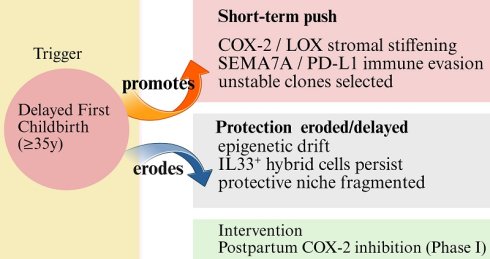
Delayed first childbirth (35-39 y) is a rapidly rising global exposure that reshapes postpartum breast cancer (PPBC) risk in a biphasic manner: a prolonged short-term risk peak (hazard ratio 1.40 at 6.4 years postpartum) coupled with long-term protection delayed beyond 15 years. Synthesizing 15 prospective cohorts with single-cell and spatial atlases of the aging parous mammary gland, we propose that maternal age is a dual modifier of PPBC risk. First, advanced age amplifies a pro-tumorigenic interactome encompassing COX-2/LOX-mediated stromal stiffening, SEMA7A/PD-L1-driven immune evasion, and clonal selection of genomically unstable cells. Second, age erodes the fidelity of the pregnancy-induced protective program: epigenetic drift compromises durable reprogramming and leads to incomplete clearance of IL33⁺ hybrid epithelial cells, sustaining a partially permissive tissue ecosystem. Together, a stronger short-term oncogenic push and a weaker long-term protective program account for both limbs of the biphasic curve. Its first clinical step in women aged ≥35 years at first birth should be a Phase I study restricted to safety and pharmacodynamic confirmation of COX-2/PGE₂ suppression, with efficacy evaluation deferred until these prerequisites are met. As childbearing continues to be delayed globally, mechanism-based precision prevention is essential to reducing the PPBC burden.
Keywords: postpartum breast cancer, delayed first childbirth, IL33+ hybrid epithelial cells, COX-2 inhibition, precision prevention, mammary gland involution
1. Introduction
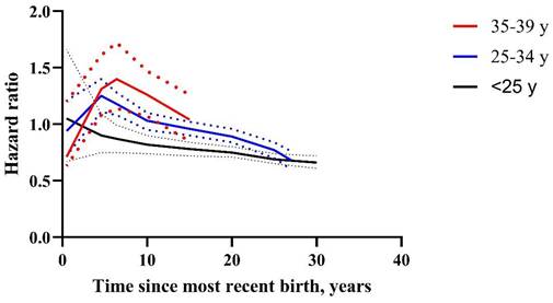
Pregnancy remodels the mammary gland across the lifespan [1, 2]. These physiological processes, when dysregulated, can create a permissive environment for oncogenesis [3, 4]. The epidemiology has never been tidy. Pregnancy protects against breast cancer in the long run, yet the first 5-10 years after delivery bring a real excess of risk [5, 6]. Clinicians have lived with that contradiction for decades, and the part that stays hardest to explain is where maternal age sits in the balance [6]. Population-based pooled analyses involving 15 prospective studies have firmly established that delayed first childbirth (35-39 y) reshapes risk in a biphasic, age-dependent manner: a higher and longer-lasting short-term risk peak (HR 1.40 at 6.4 years) versus minimal transient risk and earlier onset of protection for women with first birth before age 25 y [6]. The protective transition in women with delayed first childbirth extends beyond 15 years [6]. This stark contrast underscores that maternal age fundamentally reshapes the mammary gland's response to pregnancy, yet the underlying mechanisms remain incompletely defined [3-6]. Figure 1 plots the three trajectories side by side.
Hazard ratio for breast cancer stratified by age at first birth. Red line (35-39 y): peak HR 1.40 (95% CI 1.14-1.72) at 6.4 years since most recent birth; risk elevation persists more than 15 years. Blue line (25-34 y): transient peak; crossover to protection within 10 years. Black line (<25 y): no early increase was apparent, and the hazard ratio stayed at or below 1.0 from the earliest interval onward. The figure was created by GraphPad Prism 8. Data extracted from the discrete published key points of Nichols HB et al. Ann Intern Med 2019.
Postpartum breast cancer (PPBC) is now formally defined by international consensus as invasive carcinoma diagnosed within 10 years of childbirth [7]. It behaves differently from breast cancer in non-postpartum women: the tumors proliferate faster, the immune response is more suppressed, the genomes are less stable, and outcomes are worse [8, 9]. Incidence is climbing as childbearing age climbs [10], and PPBC is unevenly spread across subtypes, with triple-negative and HER2-positive disease over-represented, the two subtypes that respond least well to standard therapy [8, 9]. Recent advances in single-cell genomics and spatial transcriptomics have revealed unprecedented cellular and molecular dynamics within the aging and parous mammary gland [11-13]. Olander et al. used that approach to find a rare population of interleukin 33-positive (IL33+) hybrid epithelial cells: they build up with age in nulliparous females, and an early pregnancy clears them efficiently [14]. We propose that delayed first childbirth not only amplifies pro-tumorigenic signaling cascades but also impairs the fidelity of pregnancy-induced protective reprogramming, leading to incomplete clearance of this risk substrate and sustained vulnerability to breast carcinogenesis [14].
Here, we synthesize population-level epidemiological data, single-cell atlases, and mechanistic preclinical evidence to explain how advanced maternal age reshapes PPBC risk. We outline the age-potentiated pro-tumorigenic network, then characterize defective execution of protective programs, and finally translate these insights into a roadmap for biomarker development and precision prevention, focusing on tractable interventions.
2. Methods
We searched PubMed, Web of Science, and Scopus from inception to June 30, 2026, without language restrictions. The search combined controlled vocabulary and free-text terms in three thematic blocks: (i) population and exposure: “postpartum breast cancer”, “parity”, “age at first birth”, “delayed childbearing”, and “advanced maternal age”; (ii) mammary biology: “mammary gland involution”, “post-lactational regression”, “mammary stem cell”, “single-cell RNA sequencing”, “spatial transcriptomic”, “epigenetic reprogramming”, and “extracellular matrix”; and (iii) “mechanism and intervention”. Reference lists of eligible articles and recent narrative reviews were manually screened to identify additional studies. Search terms were combined systematically using Boolean operators (OR within each thematic block, AND between blocks), and records retrieved from the three databases were de-duplicated using citation management software before screening.
Because this is a hypothesis-driven narrative review rather than a systematic review, we applied explicit, framework-guided prioritization criteria instead of formal study selection. We prioritized large prospective cohorts, meta-analyses, genome-wide studies, and single-cell transcriptomic analyses, followed by functional in vivo studies using genetic or pharmacological perturbation; in vitro or correlative human studies were used only when higher-tier evidence was unavailable and are flagged as such in the text. Within each tier, studies of postpartum involution biology were preferentially included. Murine findings were retained only where directly relevant human data are absent, and these species limitations are explicitly identified. This narrative review did not perform a formal risk-of-bias assessment or meta-analysis.
3. The age-potentiated pro-tumorigenic interactome
Why is the short-term risk worse in older primiparous women? Not because one pathway switches on, but because several reinforce one another, and maternal age amplifies all of them. We group the network into three layers: stromal remodeling, immune evasion operating at several levels, and selection of genomically unstable clones.
3.1 Stromal remodeling and extracellular matrix stiffening
Elevated TGF-β1 signaling drives robust activation of lysyl oxidase (LOX) in involution-activated fibroblasts [15]. LOX cross-links collagen, and the excess cross-linking stiffens the extracellular matrix (ECM) [16]. The mechanically rigid microenvironment promotes tumor cell invasion, survival, and progression through discoidin domain receptor 2 (DDR2) and insulin-like growth factor signaling pathways [15, 17]. This process is further amplified in aged tissues by the accumulation of senescent stromal cells and a senescence-associated secretory phenotype (SASP) characterized by high levels of interleukin-6 (IL-6), matrix metalloproteinase-3, and chemokine ligand 1 [18, 19].
In vivo transplantation of involution-derived fibroblasts speeds tumor growth and reduces intra-tumoral CD8⁺ T-cell infiltration, and raises the number of lymphocyte antigen 6C-positive myeloid cells [15]. Clinically, collagen alignment in ductal carcinoma in situ (DCIS) specimens was positively associated with recurrence (HR = 1.25 per interquartile range increase in average fiber angles; 95% CI, 0.84-1.87), although this association did not reach statistical significance because the 95% CI crosses 1.0, with collagen fibers perpendicular to the duct perimeter more frequent in lesions with poor prognostic features [20]. Parity itself changes the matrix. Compared with nulliparous tissue, parous rat and human breast matrix contains more collagen I but is less linearized and less stiff [21]. DDR2 engagement by collagen promotes tumor cell invasion, and pharmacological disruption of DDR2-collagen interactions with small molecule inhibitors has been shown to blunt breast cancer metastasis in preclinical models [22]. Involution additionally licenses epithelial-mesenchymal transition, as lactogenic-hormone withdrawal during post-lactational regression induces EMT in regressing mammary epithelium [23].
3.2 Multi-layered immune evasion
Immune suppression during postpartum involution is physiological, which can be further amplified by age-related changes in the mammary microenvironment [24]. Elder and colleagues showed that semaphorin-7A (SEMA7A) promotes programmed death-ligand 1 (PD-L1) expression in immune cells of the mammary tissue during involution [24]. SEMA7A engages integrin α1β1 and transcriptionally upregulates PD-L1, impairing CD8⁺ T-cell infiltration, cytotoxicity, and immune surveillance [24]. Importantly, aging itself has been shown to directly induce SEMA7A expression in mammary tissue, suggesting that the aged gland may be primed for a stronger PD-L1 response upon involution [25]. Sherman and colleagues independently confirmed that postpartum breast cancer exhibits a distinct T cell exhaustion signature, providing orthogonal validation of this immune evasion phenotype [8].
The transient upregulation of SEMA7A during postpartum involution is a conserved physiological event [24]. In young mammary glands, this SEMA7A-PD-L1-driven window is brief and is effectively “closed” by the subsequent protective program [24, 26]. However, the aged gland closes that window badly. It resets its epigenome less faithfully [26], it carries an expanded luminal progenitor compartment with telomere dysfunction [27], and its adaptive immune education is blunted [28]. The transition into protection becomes slow and incomplete.
Beyond the SEMA7A/PD-L1 axis, additional immunosuppressive layers operate in the postpartum gland [29, 30]. Chemokine (C-C motif) ligand 8 (CCL8) is overexpressed during involution, driving the recruitment of immunosuppressive M2-type tumor-associated macrophages [29]. Neutralizing CCL8 in mouse models decreases F4/80-positive CD206⁺ macrophages and impairs lung metastasis [29]. At the same myeloid layer, C-X-C motif chemokine ligand 12 (CXCL12), the most highly expressed chemokine in these fibroblasts, recruits Ly6C⁺ monocytes to the tumor border and is associated with reduced intratumoral CD8⁺ T cell infiltration in vivo [15]. Wu and colleagues recently identified that mobilizing antigen-presenting mast cells can overcome anti-PD-1 resistance in triple-negative breast cancer, suggesting that mast cell-targeted strategies may have therapeutic potential [30]. Together, these pathways establish a multi-layered immune-evasive microenvironment that enables early tumor development and progression, particularly in triple-negative and HER2-positive subtypes.
Murine involution models carry most of the weight for the SEMA7A/PD-L1 axis [24]. Where human tissue enters the picture the support narrows: T-cell exhaustion signatures have been checked in PPBC specimen [8], and little beyond that. Serial human postpartum biopsies across maternal ages are also missing, and the age-dependent dynamics of SEMA7A induction have been demonstrated in mouse but not human tissue [25]. CCL8 and M2 macrophages have been characterized exclusively in mice [29]. These species-specific limitations should be considered when interpreting the proposed immune evasion network.
3.3 Hypoxia and clonal selection of genomically unstable clones
Recent evidence implicates intratumoral hypoxia as a potential driver of PPBC aggressiveness [31]. Bakhuis et al. analyzed tumor tissues from 148 pregnancy-associated breast cancer cases diagnosed during pregnancy and 45 PPBC patients, and found that hypoxia-related immunohistochemical markers are frequently expressed and are associated with worse prognosis in both pregnancy-related breast cancer (diagnosed during gestation) and postpartum cases [31]. Because the analysis relied on static immunohistochemical markers in archival tissue, it supports a clinical prognostic association rather than clonal evolutionary dynamics or genomic instability; we therefore treat hypoxia marker expression as a correlate of aggressive clinical behavior, not as evidence of active clonal selection. Here we emphasize that PABC encompasses tumors diagnosed during active gestation, whereas PPBC refers to tumors diagnosed after delivery, during or after postpartum involution [3, 7]: Lyons et al. proposed that these be regarded as two distinct subtypes, whereas Amant et al. have argued more radically that the umbrella term PABC is outdated and should no longer be used [3, 7]. The systemic endocrine and immune landscapes of these two windows differ fundamentally, with gestation characterized by increased exposure to estrogen and progesterone together with the immunosuppressive effects of pregnancy, and involution characterized by widespread epithelial apoptosis and inflammatory remodeling [3]. Mixed PABC cohorts should therefore be interpreted as approximations of, rather than direct evidence for, involution-specific PPBC biology, and studies that disaggregate gestational from postpartum diagnoses will be required to isolate involution-specific mechanisms [7, 31].
The hypoxic, inflammatory, and mechanically stressed microenvironment described above imposes strong selective pressure, consistent with the hypothesis that such microenvironmental pressures may select for genomically unstable clones. Direct support for a distinct postpartum biology comes from transcriptomic profiling: using RNA expression data from clinically matched estrogen receptor (ER)-positive cases (n = 16), Jindal et al. showed that breast cancers in young women can be distinguished on the basis of a postpartum versus nulliparous diagnosis, with PPBC signatures indicating increased cell-cycle activity and T-cell activation together with reduced estrogen receptor and TP53 signaling [9]. When this signature was applied to a validation cohort of young women's breast cancer (n = 311), high scores were associated with a significantly reduced 15-year overall survival (HR 2.13, P = 0.0011), indicating that PPBC is a molecularly distinct entity with poor-prognosis phenotypes [9]. Because the comparison was made against nulliparous rather than gestational cases, it avoids the diagnostic mixing that limits other PABC cohorts [9]; nevertheless, larger sequencing studies that separate gestational from postpartum diagnoses are needed before anyone can describe the mutation pattern that belongs specifically to the involution window. These data are consistent with, but do not prove, the hypothesis that the postpartum microenvironment selectively amplifies genomically unstable clones and thereby transiently increases breast cancer risk [9].
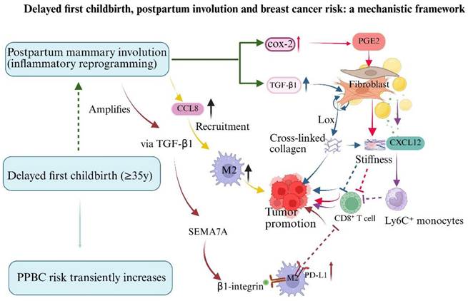
Clinical evidence further underscores the aggressive nature of PPBC. Al-Masri et al. found that HER2-positive pregnancy-associated breast cancer patients have significantly worse recurrence-free survival compared to non-pregnancy-associated breast cancer controls (P = 0.026), with PPBC patients faring particularly poorly [32]. Their cohort mixed tumors diagnosed during pregnancy with postpartum tumors, so the result should not be carried over to strictly defined PPBC without qualification [32]. Notably, pregnancy-associated breast cancer patients experienced significant delays in initiating anti-HER2 therapy (mean 21.3 weeks vs 16.9 weeks, P = 0.02), suggesting that treatment delay may contribute to the worse outcomes observed in this population [32]. Collectively, these interconnected pathways are summarized schematically in Figure 2. A central unresolved question is whether delayed first childbirth amplifies postpartum mammary involution through age-related inflammatory aging (dashed arrow in Figure 2). Addressing this will require comparing involution markers, such as COX-2, TGF-β1, NF-κB, stromal stiffening, and immunosuppressive infiltrate, between delayed (≥35 y) and young primiparas; testing age at first birth as an effect modifier of postpartum risk in large prospective cohorts; and using timed-primiparity and senolytic models to establish causality. Importantly, uncertainty about this amplifier does not weaken the two core pathways summarized in Figure 2, because those pathways are each supported by evidence independent of maternal age.
Delayed first childbirth, postpartum mammary involution, and breast cancer risk: a mechanistic framework. Delayed first childbirth (≥35 years) is associated with a transient increase in postpartum breast cancer (PPBC) risk [6]. Postpartum mammary involution drives an age-independent tumor-promoting microenvironment through multiple branches: (i) COX-2-derived PGE2 cooperates with TGF-β1 to activate fibroblasts, leading to lysyl oxidase (LOX)-mediated collagen cross-linking and stromal stiffening, which in turn suppresses CD8⁺ T cells to further promote tumor formation [15]; (ii) CCL8 recruits M2 macrophages [29]; (iii) TGF-β1 induces semaphorin 7A (SEMA7A) expression, which signals via β1-integrin and cooperatively drives PD-L1-mediated immunosuppression [24, 25]; and (iv) CXCL12 recruits Ly6C⁺ monocytes, excluding CD8⁺ T cells from the tumor microenvironment [15]. The dashed green arrow denotes a hypothesized amplifier: age-related changes in the mammary gland may amplify the involution response [37, 43], although direct evidence that delayed childbirth per se exacerbates involution is lacking. This uncertainty is confined to the amplifier link and does not downgrade the established epidemiologic association or the age-independent involution biology. The figure was created in BioRender.
4. The impaired fidelity of the protective program: How Delayed First Childbirth Attenuates Pregnancy-Induced Breast Cancer Protection
4.1 Epidemiological foundation: protection delayed and attenuated
Epidemiological evidence clearly establishes that delayed first childbirth attenuates pregnancy-induced breast cancer protection. In the pooled analysis of 15 prospective cohorts by Nichols et al., women with first birth at ≥35 years reached a peak hazard ratio of 1.40 (95% CI 1.14-1.72) at 6.4 years postpartum, and did not transition to protective status until more than 15 years after delivery [6]. Albrektsen et al. independently confirmed that each 5-year delay in first birth is associated with a significantly higher and more prolonged peak risk [33]. This age-dependent degradation of protection implies that the parity-induced protective program, while initiated by pregnancy, fails to achieve full functional maturity in older primiparous women, a phenomenon that cannot be explained simply by delayed timing, but rather by fundamental impairment in the program's execution fidelity.
4.2 Incomplete epigenetic reprogramming: diminished plasticity in the aged mammary epithelium
Pregnancy induces widespread and durable epigenetic reprogramming of the mammary epithelium, establishing a stable tumor-suppressive landscape [26]. Feigman and colleagues showed that pregnancy remodels enhancer chromatin in mouse mammary epithelial cells, inducing lasting epigenetic changes that protect against cMYC-driven tumorigenesis [26]. However, the capacity for such reprogramming is fundamentally dependent on the epigenetic plasticity of the target tissue. Angarola et al. recently showed, through comprehensive single-cell aging atlases, that the healthy mammary epithelium accumulates age-associated epigenomic and transcriptomic alterations, a state termed epigenetic drift, which constrains its capacity for adaptive remodeling in response to physiological stimuli [13]. Fane and Weeraratna further established that aging-associated epigenetic rigidity, driven in part by altered Polycomb group protein activity and progressive DNA methylation changes, impairs tissue-level responses to hormonal cues across multiple organ systems [19]. In the context of delayed first childbirth, the aged mammary epithelium enters pregnancy with pre-existing epigenetic alterations that diminish both the fidelity and dynamic range of pregnancy-induced reprogramming [13, 19]. We therefore hypothesize that the installation of a complete protective epigenetic signature is incomplete, leaving residual molecular vulnerabilities that undermine long-term cancer resistance [26]. In breast tumors, an elevated PR-A/PR-B ratio is independently associated with poor prognosis [34]. Independent studies have linked age at menarche and age at first birth to PR-A promoter methylation in normal breast tissue [35, 36], suggesting that an elevated ratio may reflect age-associated epigenetic alterations. Human data further indicate that maternal age and parity remodel the mammary epithelial cell compartment directly [37].
4.3 Incomplete clearance of age-accumulated risk substrate: the IL33⁺ hybrid cell reservoir
A cornerstone of parity-induced protection is the long-term functional reduction of the mammary stem cell (MaSC) pool, the putative cells of origin for many breast cancers [38, 39]. Siwko et al. showed that early pregnancy causes a persistent decrease in functional mammary epithelial stem cells, which simply leaves fewer cells available to transform [38]. Meier-Abt et al. subsequently revealed a mechanistic basis for this stem cell “lockdown”: early pregnancy induces terminal differentiation of basal stem/progenitor cells and downregulates the Wnt/Notch signaling ratio, thereby reducing their proliferative potential and clonal expansion capacity [39]. Rocha et al. recently extended this framework by demonstrating that loss of luminal receptor activator of nuclear factor-κB signaling in parous mammary glands reduces cell fitness and drives basal cell bipotency, shifting the differentiation program once more [40].
The discovery by Olander et al. provides a compelling cellular paradigm for how delayed first childbirth may compromise this protective mechanism [14]. Through single-cell transcriptomic analysis of nulliparous and parous mammary glands across the murine lifespan, they found a rare population of IL33+ hybrid epithelial cells co-expressing basal and luminal markers that accumulates preferentially in the aged nulliparous mammary gland but is profoundly diminished in age-matched parous counterparts [14]. Functionally, IL33 treatment recapitulated aged nulliparous phenotypes, including basal cell expansion and increased organoid formation, which was further potentiated by Trp53 knockdown [14]. In aged nulliparous mice the population keeps expanding, whereas an early pregnancy clears it, apparently through differentiation-inducing signals [14]. We hypothesize that in delayed first childbirth (≥35 years), the aged gland's diminished epigenetic receptivity and impaired differentiation signaling may result in incomplete clearance of this IL33+ population. This hypothesis is based on murine data; direct evidence from human postpartum tissue is currently lacking. These persisting hybrid cells may serve as a residual reservoir of transformation-prone cells, directly contributing to sustained, long-term risk [14]. Two lines of evidence support the idea: an early pregnancy is a strong driver of MaSC depletion and epigenetic maturation [38, 39]; while the aging gland independently expands stem/progenitor-like compartments and differentiates less readily [13, 27]. The IL33+ hybrid cell represents a newly identified component of this residual reservoir that may escape the protective lockdown program [14].
4.4 A fragmented tumor-suppressive microenvironment
The establishment of a tumor-suppressive microenvironment after pregnancy depends on three interdependent components: normalized stromal architecture [21], durable immune surveillance [28, 41], and favorable metabolic adaptation [42]. Age interferes with all three. What results is not a barrier but a patchwork, with gaps a developing tumor can use [19, 26].
4.4.1 Fibrosis propensity
Pregnancy induces lasting alterations in mammary stromal architecture that contribute to the protective phenotype. Maller et al. showed that parity changes mammary ECM composition and organization, parous matrix has more collagen I but is less linearized and less stiff than nulliparous matrix [21]. This shift toward a more randomized collagen fiber organization reduces local tissue stiffness and mechanically discourages tumor cell invasion [21]. The clinical counterpart is that aligned collagen perpendicular to the duct perimeter tracks with poor prognostic features in DCIS and with a non-significant trend toward recurrence, as detailed in Section 3.1 [20]. However, the aged mammary stroma exhibits a heightened baseline propensity for fibrosis and aberrant ECM deposition, driven by accumulation of senescent stromal cells and a SASP [18, 19]. The SASP, which comprises a complex mixture of pro-inflammatory cytokines, chemokines, and matrix metalloproteinases [18], promotes excessive collagen cross-linking and tissue stiffening. This age-acquired profibrotic state likely impairs the extent and efficiency of pregnancy-induced stromal normalization, potentially resulting in a residual pro-invasive mechanical microenvironment [19, 20]. Lyons et al. showed the same biology from the disease side: postpartum LOX-mediated collagen cross-linking drives DCIS toward invasive carcinoma [16]. The stromal compartment also modifies how age and risk exposures shape age-related lobular involution dynamics [43].
4.4.2 Immunosenescence
The postpartum period involves significant immune activation and remodeling. Over the long term, pregnancy is considered an immune-educating event, potentially establishing a population of tissue-resident memory T cells that provide durable surveillance [41]. Furthermore, the mammary macrophage population shifts from the pro-tumorigenic M2-like phenotype prevalent during early postpartum to a predominance of antitumor M1-like macrophages in the parous state [44]. Hitchcock et al. showed that the immune environment shifts throughout post-lactational regression, with changes in macrophage polarization that correlate with tumor growth rate [45]. However, aging fundamentally alters immune competence through immunosenescence, a progressive deterioration of both innate and adaptive immune functions [28]. Nikolich-Žugich found that aging impairs T-cell repertoire diversity, reduces naive T-cell output, and blunts the capacity for de novo immune responses [28]. In the mammary gland specifically, Kines et al. found that aging induces SEMA7A expression, which promotes TGF-β1-mediated epithelial cell plasticity and breast tumor metastasis, indicating that the aged mammary immune microenvironment is primed toward immune tolerance [25]. These age-related immune deficits likely impair the establishment of a robust, long-lived immune surveillance network following pregnancy in older primiparous women, permitting a more permissive immune landscape [25].
4.4.3 Inflammaging
Parity induces beneficial systemic metabolic changes that contribute to the protective phenotype. As reviewed by Iyengar et al., reproductive history can modulate adipokine secretion profiles; an increased adiponectin-to-leptin ratio, for instance, has been associated with reduced local inflammation and epithelial proliferation [46]. Grossmann and Cleary established the mechanism in preclinical models: leptin pushes proliferation and inflammation, adiponectin opposes both, and the balance between them modulates mammary tumorigenesis directly [47]. However, aging is associated with decreased metabolic flexibility and a shift toward a chronic, low-grade inflammatory state termed “inflammaging” by Franceschi et al., characterized by elevated circulating pro-inflammatory cytokines, altered adipokine profiles, and metabolic dysregulation [42]. This age-associated inflammaging milieu creates a systemic environment that is fundamentally different from the metabolic context in which pregnancy occurs at a young age [42]. The interaction between late first pregnancy and an aged systemic metabolic milieu may therefore attenuate these beneficial adaptations, sustaining a more pro-proliferative and inflammatory environment conducive to oncogenesis [42, 46].
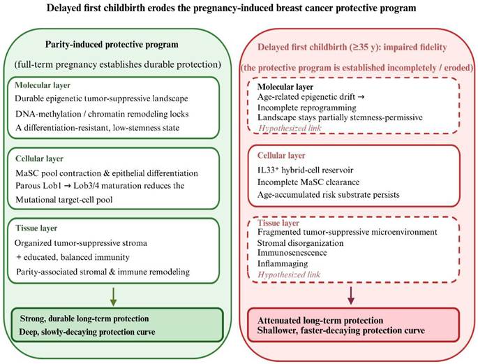
We propose that delayed first childbirth erodes the protective program at three levels in sequence. Molecularly, age-related epigenetic drift prevents the stable installation of tumor-suppressive chromatin marks [13, 26]. Cellularly, the aged niche loses its differentiation-inducing capacity, resulting in incomplete clearance of IL33⁺ hybrid cells and an incompletely depleted mammary stem cell pool [14, 38]. At the tissue ecosystem level, stromal normalization is impaired [19], immune education remains blunted [28], and metabolic adaptation is attenuated [42]. Each failure is partial, and together they stop a coherent tumor-suppressive microenvironment from assembling. Pregnancy does initiate the program, but in an older primipara it never reaches full maturity, and that is what we think produces protection that is both weaker and later [6, 48]. This conceptualization of protection as a modifiable developmental outcome with variable fidelity, dependent on both the quality of the initiating pregnancy signal and the receptive competence of the target tissue, reframes the clinical challenge from passive risk observation to the potential for active intervention [49]. Figure 3 sets the three levels out side by side, contrasting the fully executed program of an early full-term pregnancy with the partly installed version after delayed first childbirth, and marking established links solid and hypothesized ones dashed.
Delayed first childbirth erodes the pregnancy-induced breast cancer protective program. Left: components induced by a full-term pregnancy. Right: their impairment under delayed first childbirth. Left (parity-induced protective program): the molecular layer reflects the durable epigenetic tumor-suppressive landscape established by pregnancy [26]; the cellular layer reflects parity-induced differentiation and contraction of the mammary stem/progenitor cell pool [39, 40]; and the tissue layer reflects organized, tumor-suppressive stroma together with parity-associated immune remodeling [21, 41]. Right (delayed first childbirth, ≥35 years): age-related epigenetic drift with incomplete reprogramming [13, 19]; persistence of an IL33⁺ hybrid-cell reservoir with incomplete clearance of the mammary stem cell compartment [14]; and a fragmented tumor-suppressive microenvironment comprising stromal disorganization, immunosenescence, and inflammatory aging [13, 21]. Elements shown in italics are hypothesized links that await direct testing. The figure was created in BioRender.
5. Risk stratification and candidate biomarkers
5.1 Clinical stratification
Clinical factors associated with elevated postpartum risk include age ≥35 years at first birth, rapid or dysregulated post-weaning involution. Additionally, preoperative Ki-67 levels have also been proposed as a candidate marker, although the evidence for an independent association remains limited [6, 9]. These factors provide a practical framework for initial risk stratification in clinical settings. Nichols et al. further showed that both the timing of risk peak and the duration of risk elevation are directly correlated with maternal age, providing a simple clinical tool for risk assessment [6].
5.2 Molecular and genomic biomarkers
Candidate molecular biomarkers for risk stratification span multiple modalities. At the epigenetic level, the progesterone receptor isoform A/ progesterone receptor isoform B (PR-A/PR-B) protein ratio measured in breast tissue or nipple aspirate fluid could serve as an indicator of successful reprogramming [34, 35]. Circulating COX-2 metabolites, such as PGE₂ or its stable degradation products, may reflect the degree of inflammatory activation during involution [15, 16]. At the cellular level, the burden of IL33+ hybrid epithelial cells, if detectable in breast biopsies or nipple aspirate fluid, could provide a direct readout of residual nulliparous aging signatures in women with delayed first childbirth [14]. However, this marker remains entirely preclinical: IL33+ hybrid epithelial cells have been characterized only in murine tissue, and neither their existence in the human postpartum gland nor their detectability in human specimens has been demonstrated; they should therefore be regarded as an investigational hypothesis rather than a biomarker with clinical readiness, pending validation in human postpartum tissue. SEMA7A expression levels in breast tissue or serum may correlate with the degree of immune evasion [24]. It should be noted that the age-dependent SEMA7A/PD-L1 dynamics underlying this proposal have been demonstrated in mouse models but not yet in serial human postpartum samples [24]. Hypoxia-related immunohistochemical markers reported by Bakhuis et al., may add to risk stratification [31]. TP53 mutation status and MYC amplification, detectable through liquid biopsy approaches, may identify women at highest risk for aggressive PPBC subtypes [50]. Circulating tumor DNA (ctDNA) is the more speculative option: Saura and colleagues detected ctDNA in the breast milk of women with PPBC up to 18 months before clinical diagnosis, which suggests that monitoring during the postpartum period could catch clonal expansion early, though the approach is still investigational [51].
6. Preventive strategies: mechanism-informed interventions
Viewing protection as a modifiable developmental outcome with variable fidelity redirects clinical priorities. Short-term risk still matters, but the more interesting question is whether we can push the protective program to completion in the women least likely to complete it on their own.
6.1 Short-term postpartum COX-2 inhibition: rationale and safety considerations
COX-2 is the most druggable node in the postpartum pro-tumorigenic niche [15]. Preclinical models demonstrate that short-term postpartum administration of selective COX-2 inhibitors suppresses inflammation, stromal stiffening, and immune suppression, thereby reducing tumor promotion [15]. Ibuprofen given during involution restores anti-tumor immunity and delays outgrowth in mice [15]. Functional human validation of the COX-2/PGE₂ axis in the postpartum window remains limited [16]; the optimal therapeutic window should be established by dedicated pharmacokinetic and pharmacodynamic studies rather than assumed from preclinical models.
This strategy is supported by substantial biological rationale; but translating it needs more caution than the preclinical literature invites. Involution is a physiological process, not a disease, and it needs inflammatory cytokines to clear differentiated epithelial cells by apoptosis, to remodel tissue, and to maintain the antimicrobial barrier that protects against mastitis [45, 49]. Damping those pathways in a healthy postpartum woman risks interfering with normal remodeling, and a preventive agent has to clear lactation safety thresholds and regulatory requirements considerably stricter than those applied to treating established disease [49]. With these constraints in mind, the immediate priority is a Phase I trial to establish safety, mammary-compartment pharmacokinetics and pharmacodynamic suppression of the COX-2/PGE₂ axis rather than an efficacy-oriented Phase I/II trial before this strategy can be advanced in women aged ≥35 years at first childbirth [15, 49]. However, before clinical translation, three principal safety concerns must be addressed, concerning the involuting mammary gland, the infant in the settings where exposure remains possible, and maternal cardiovascular health. First, mammary-compartment drug exposure and infant safety. Celecoxib, the best-studied selective COX-2 inhibitor in lactation, has a milk-to-plasma ratio of 0.23 (95% CI: 0.15-0.31) and a relative infant dose of approximately 0.3% of the maternal weight-adjusted dose—well below the 10% safety threshold [52, 53]. No adverse effects have been reported in breastfed infants of mothers taking celecoxib 200 mg daily, and both the Association of Anaesthetists and EULAR consider celecoxib compatible with breastfeeding [54]. It should be noted, however, that the EULAR recommendations address the use of antirheumatic drugs in patients with rheumatic disease rather than chemoprevention in healthy women; the risk-benefit tolerance for a preventive indication in a healthy postpartum population is fundamentally stricter, so this compatibility classification informs, but does not by itself sanction, a preventive use of celecoxib.
Second, potential interference with involution. COX-2-dependent inflammatory signaling is a physiological component of mammary involution [15]. Complete pharmacological suppression could theoretically delay glandular regression or cause unresolved engorgement. Beyond glandular regression, inflammatory mediators generated during involution also coordinate apoptotic cell clearance and extracellular matrix remodeling, and innate immune defenses of the involuting gland contribute to protection against mastitis [45, 49]. These biological trade-offs must be explicitly monitored in any early-phase study, with predefined endpoints including the kinetics of glandular regression, lactation outcomes after treatment, and the incidence of postpartum mastitis. However, preclinical studies indicate that short-term, low-dose COX-2 inhibition reduces tumor-promoting inflammation without ablating the involution process itself [15, 49].
Third, cardiovascular safety. A meta-analysis confirmed that celecoxib does not significantly increase cardiovascular events compared with placebo and may reduce all-cause mortality compared with non-selective NSAIDs [55]. Critically, cardiovascular risk is dose-dependent: at 400 mg once daily, the hazard ratio is 1.1 (95% CI 0.6-2.0); at 200 mg twice daily, 1.8 (95% CI 1.1-3.1); and at 400 mg twice daily, 3.1 (95% CI 1.5-6.1) [56]. The proposed short-term (days to weeks), low-dose regimen in young, otherwise healthy postpartum women represents a substantially lower cardiovascular risk profile than the chronic, high-dose regimens evaluated in elderly arthritis populations. Nevertheless, baseline cardiovascular assessment and exclusion of women with active cardiovascular disease are essential.
In summary, the safety profile of short-term, low-dose celecoxib in the postpartum setting appears favorable based on existing lactation pharmacokinetic data and cardiovascular safety trials, but dedicated Phase I studies in postpartum women are mandatory before proceeding to efficacy trials.
Finally, even a favorable safety profile would not by itself enable implementation, because chemoprevention in healthy postpartum women faces regulatory hurdles that are distinct from those of therapeutic trials. Because breast secretion may still be expressed for engorgement relief and subsequently fed or donated, any such product must carry an explicit instruction on the discard of expressed milk and on the washout interval before feeding or donation; placebo-controlled designs raise ethical considerations in breastfeeding populations; and the modest absolute short-term incidence of PPBC means that early-phase trials must rely on pharmacodynamic biomarkers and intermediate endpoints rather than on cancer incidence, which is far too rare for adequately powered prevention trials within this window. Regulators will want to see on-target pathway modulation and formal lactation safety data before efficacy can even be discussed. So, we position short-term postpartum COX-2 inhibition as a strategy with solid biological grounding that is still investigational: the first clinical step is a dedicated Phase I safety and pharmacodynamic study run with involution monitoring in place, not a Phase I/II efficacy trial.
6.2 Sequential targeted interventions
Women selected as high risk by biomarker profile could then move on to sequential strategies. LOX inhibitors are one option for persistent ECM stiffening [16, 57]. LOX-mediated collagen cross-linking drives fibrosis-enhanced metastasis in several cancer types [57]. For women with residual M2 macrophage infiltration, targeting the CCL8/CCR2 axis may be explored [29]. These approaches remain investigational and require further clinical validation in prospective trials. A phased research framework that prioritizes short-term COX-2 inhibition as the foundational step, followed by biomarker-guided sequential combinations and, where appropriate, therapeutic immune checkpoint blockade, is outlined in Table 1.
A risk-stratified research agenda for postpartum breast cancer prevention
| Proposed Risk Stratum | Biomarker Enrichment Strategy | Prioritized Investigational Approach |
|---|---|---|
| Stratum A (Foundational Study) | Age ≥ 35 years and/or elevated stromal remodeling (imaging/biomarker) | Phase I safety and pharmacodynamic trial of postpartum COX-2 inhibition Goal: Establish safety and on-target biological activity before any efficacy evaluation |
| Stratum B (Next Phase Targets) | Evidence of active fibrotic/immunosuppressive pathways despite COX-2 inhibition | Preclinical evaluation of LOX or CCR2 inhibitors to the foundational regimen Goal: Evaluate dual-pathway inhibition |
| Stratum C (Therapeutic Context Only) | Postpartum TNBC with high PD-L1 expression | Restrict immune checkpoint blockade to treatment trials Goal: Improve outcomes for established disease |
6.3 Immune and metabolic interventions
Immune checkpoint inhibitors are not suitable for preventive applications due to their safety profile in healthy individuals [58]. The known immune-related adverse event profile of checkpoint inhibitors, including pneumonitis, colitis, and endocrinopathies, precludes their use for primary prevention in healthy women, where the risk-benefit ratio is unfavorable [58]. Their evaluation should remain strictly within the domain of therapeutic trials for established PPBC, particularly for triple-negative and HER2-positive subtypes with high PD-L1 expression [24, 58].
Metabolic interventions such as lifestyle change and weight management could help at the margins by lowering systemic inflammation and improving metabolic flexibility [42, 46, 47, 59]. However, prospective evaluation is required before clinical implementation in the postpartum setting.
7. Future research priorities
The field of postpartum breast cancer research faces several critical gaps that must be addressed to translate mechanistic insights into clinical practice:
1. Age-stratified single-cell and spatial transcriptomic atlases. The lack of age-stratified single-cell atlases of the human postpartum mammary gland represents a major barrier to understanding how maternal age modulates cellular responses to pregnancy [13, 14]. Multi-institutional efforts are needed.
2. Animal models recapitulating delayed first pregnancy. No existing model reproduces delayed first pregnancy while allowing lifetime cancer risk to be measured, which caps what mechanistic work can do [19, 60]. Age-controlled murine models across the lifespan are needed.
3. Prospective clinical trials of COX-2 inhibition. No registered trial targets involution biology in older primiparous women, and that is a gap which could be closed now [49]. The first such study should be a Phase I trial establishing safety, tolerability, and pharmacodynamic biomarkers of COX-2 pathway suppression, without formal efficacy endpoints, and it should build in involution monitoring from the start, covering the kinetics of glandular regression and the incidence of mastitis, before any efficacy-oriented evaluation follows.
4. Biomarker validation studies. PR-A/PR-B ratio, IL33+ cell burden, hypoxia markers and collagen alignment scores all need large-scale cohort testing as independent predictors of PPBC risk [20, 31, 34], with longitudinal biosampling across the peripartum period so that the natural history of each substrate can be described.
5. Liquid biopsy and radiomics development. Non-invasive approaches hold promise for risk stratification in postpartum populations. For liquid biopsy, emerging evidence suggests that ctDNA monitoring could detect early clonal expansion during the postpartum period. Notably, Saura and colleagues demonstrated that ctDNA is detectable in the breast milk of women with postpartum breast cancer up to 18 months prior to clinical diagnosis, offering a promising avenue for early detection [51]. For imaging, MRI-based radiomic signatures, particularly quantitative background parenchymal enhancement, have been independently associated with increased breast cancer risk and need further validation in postpartum populations [61, 62].
6. Mechanistic studies of protective program restoration. Proof-of-concept studies are needed to determine whether targeted interventions, such as LOX inhibitors or CCR2 antagonists, can restore a tumor-suppressive state in high-risk cohorts after the postpartum window has closed [29, 57]. How long that window stays open is a design question, and it needs an answer before trials are built.
7. International collaborative initiatives. Filling these voids will require prospective, multi-institutional initiatives that integrate longitudinal biosampling, advanced imaging, and mechanistic intervention arms. Such efforts should be prioritized given the rapid global shift toward delayed childbearing [10, 63].
8. Limitations and knowledge gaps
Several limitations bound how far this framework can be carried. First, limited human mechanistic evidence. Most data derive from murine models or cell lines. The PR-A/PR-B switch has been studied mainly in breast tumors [34]; how the ratio behaves with age in normal postpartum human breast has never been recorded. The IL33⁺ hybrid epithelial cell has been characterized only in mice [14]; its existence in the human postpartum gland and correlation with age at first birth remain unproven. The SEMA7A/PD-L1 axis has been demonstrated in murine involution and human cancer specimens [24, 25], but serial human postpartum biopsies across maternal ages are lacking. Until age-stratified human cohorts are assembled, the mechanisms proposed here remain inferences. Second, incomplete confounding control. Delayed childbearing correlates with higher socioeconomic status, lower parity, obesity, metabolic syndrome and assisted reproductive technologies, each independently modulating breast cancer risk [10, 46]. Pooled analyses adjusted for several of these variables [6], but residual confounding cannot be excluded. Some risk amplification attributed to maternal age may instead reflect correlated exposures or their interactions with age. Third, additional gaps. The integrated cascade has not been experimentally validated as a whole; individual nodes have been studied in isolation. Fourth, limited population diversity and potential for selection bias in our narrative synthesis. Findings derived predominantly from European-ancestry populations may not generalize to other racial/ethnic groups given known differences in PPBC incidence and subtype distribution, parity patterns, and breastfeeding practices. The Nichols et al. pooled analysis of 15 prospective cohorts was composed primarily of North American and European populations [6], and key mechanistic studies (Olander et al. [14], Guo et al. [15], Elder et al. [24]) utilized murine models or European-derived human specimens. Asian populations merit particular attention. Asian women exhibit a higher proportion of HER2-enriched subtypes [43, 64], lower baseline estrogen levels that may alter involution dynamics, and distinct parity-breastfeeding patterns that could modulate the postpartum risk window. Finally, narrative reviews select. As a hypothesis-driven synthesis, this review prioritized evidence supporting the proposed mechanistic framework. Studies reporting null or contradictory findings, for example, cohort analyses that did not observe a transient postpartum risk elevation, or studies where age at first birth did not significantly modulate postpartum risk, were not systematically sought or appraised. The framework should therefore be considered a proposed hypothesis rather than a balanced summary of all available evidence. Future systematic reviews and meta-analyses are needed to test the framework's predictions against the totality of published data.
9. Conclusions
Delayed first childbirth fundamentally reshapes the mammary gland's response to pregnancy through a biphasic mechanism: it amplifies short-term risk and delays protection beyond 15 years, in contrast to the early-onset protection seen with first birth before age 25 [6]. This review integrates population-level data [6] with single-cell and spatial transcriptomic findings [14, 26]. We demonstrate that delayed first childbirth amplifies core risk-phase pathways, including the COX-2/LOX-driven stromal-immune crosstalk [15, 17], and SEMA7A/PD-L1-mediated immune evasion [24]. Concurrently, we propose that delayed first childbirth likely impairs the establishment of long-term protective features such as immune memory [25, 41], tumor-suppressive epigenetic reprogramming [26], and the effective lockdown of the stem cell compartment [38]. One question deserves priority: does postpartum triple-negative breast cancer with high PD-L1 represent a distinct, immune-evasive subtype that should receive immunotherapy upfront [58]? Whether delayed first childbirth contributes to that subgroup is not established. Mao et al. examined reproductive risk factors across standard clinical subtypes in general breast cancer populations and did not stratify by postpartum status or PD-L1 expression [65], so postpartum-specific estimates of age at first birth in PD-L1-positive triple-negative disease are still missing. Validating the biomarkers and targets discussed here will require large-scale cohorts [6, 20], and only then can prevention be tailored to the two things delayed first childbirth does to breast cancer risk.
By bridging mechanistic understanding with translational action, this framework paves the way for reducing PPBC burden in the growing population of women with delayed first childbirth [6]. As childbearing continues to be delayed worldwide, translating these biological insights into clinical practice will become increasingly critical to reducing the global burden of postpartum breast cancer [10].
Abbreviations
CCL8: chemokine (C-C motif) ligand 8; COX-2: cyclooxygenase-2; ctDNA: circulating tumor DNA; CXCL12: C-X-C motif chemokine ligand 12; DDR2: discoidin domain receptor 2; ECM: extracellular matrix; LOX: lysyl oxidase; MaSC: mammary stem cell; PD-L1: programmed death ligand 1; PABC: pregnancy-associated breast cancer; PPBC: postpartum breast cancer; PR-A/B: progesterone receptor isoform A/B; SASP: senescence-associated secretory phenotype; SEMA7A: semaphorin-7A; TGF-β1: transforming growth factor-β1; TNBC: triple negative breast cancer.
Acknowledgements
ChatGPT was used for language polishing. The authors reviewed and edited the content as needed and take full responsibility for the content of the published article.
Author Contributions
CHZ wrote the original draft, created illustrations, and collected the literature. WZ reviewed and edited the manuscript, providing resources. ZA supervised the study, edited and reviewed the manuscript. All authors have read and approved the final version of the manuscript.
Competing Interests
The authors have declared that no competing interest exists.
References
1. Visvader JE, Stingl J. Mammary stem cells and the differentiation hierarchy: current status and perspectives. Genes Dev. 2014;28(11):1143-58
2. Valdés-Mora F, Salomon R, Gloss BS. et al. Single-cell transcriptomics reveals involution mimicry during the specification of the basal breast cancer subtype. Cell Rep. 2021;35(2):108945
3. Lyons TR, Schedin PJ, Borges VF. Pregnancy and breast cancer: when they collide. J Mammary Gland Biol Neoplasia. 2009;14(2):87-98
4. Russo J, Moral R, Balogh GA, Mailo D, Russo IH. The protective role of pregnancy in breast cancer. Breast Cancer Res. 2005;7(3):131-42
5. Lue JC, Radisky DC. From embryogenesis to senescence: the role of mammary gland physiology in breast cancer risk. Cancers (Basel). 2025;17(5):787
6. Nichols HB, Schoemaker MJ, Cai J. et al. Breast cancer risk after recent childbirth: a pooled analysis of 15 prospective studies. Ann Intern Med. 2019;170(1):22-30
7. Amant F, Lefrère H, Borges VF. et al. The definition of pregnancy-associated breast cancer is outdated and should no longer be used. The Lancet Oncol. 2021;22(6):753-4
8. Sherman ME, Etievant L, Vierkant RA. et al. Postpartum breast cancer: evidence for a distinct phenotype. J Natl Cancer Inst. 2026;118(5):926-36
9. Jindal S, Pennock ND, Sun D. et al. Postpartum breast cancer has a distinct molecular profile that predicts poor outcomes. Nat Commun. 2021;12(1):6341
10. Londero AP, Bertozzi S, Xholli A, Cedolini C, Cagnacci A. Breast cancer and the steadily increasing maternal age: are they colliding? BMC Womens Health. 2024;24(1):286
11. Bach K, Pensa S, Grzelak M. et al. Differentiation dynamics of mammary epithelial cells revealed by single-cell RNA sequencing. Nat Commun. 2017;8(1):2128
12. Davis FM, Lloyd-Lewis B, Harris OB. et al. Single-cell lineage tracing in the mammary gland reveals stochastic clonal dispersion of stem/progenitor cell progeny. Nat Commun. 2016;7:13053
13. Angarola BL, Sharma S, Katiyar N. et al. Comprehensive single-cell aging atlas of healthy mammary tissues reveals shared epigenomic and transcriptomic signatures of aging and cancer. Nat Aging. 2025;5(1):122-43
14. Olander A, Medina P, Haro Acosta V, Kaushik S, Dijkgraaf M, Sikandar SS. Divergent aging of nulliparous and parous mammary glands reveals IL33+ hybrid epithelial cells. Nat Commun. 2026;17(1):1898
15. Guo Q, Minnier J, Burchard J, Chiotti K, Spellman P, Schedin P. Physiologically activated mammary fibroblasts promote postpartum mammary cancer. JCI Insight. 2017;2(6):e89206
16. Lyons TR, O'Brien J, Borges VF. et al. Postpartum mammary gland involution drives progression of ductal carcinoma in situ through collagen and COX-2. Nat Med. 2011;17(9):1109-15
17. Slocum E, Germain D. Collagen and PAPP-a in the etiology of postpartum breast cancer. Horm Cancer. 2019;10(4-6):137-44
18. Coppé J, Patil CK, Rodier F. et al. Senescence-associated secretory phenotypes reveal cell-nonautonomous functions of oncogenic RAS and the p53 tumor suppressor. PLoS Biol. 2008;6(12):2853-68
19. Fane M, Weeraratna AT. How the ageing microenvironment influences tumour progression. Nat Rev Cancer. 2020;20(2):89-106
20. Conklin MW, Gangnon RE, Sprague BL. et al. Collagen alignment as a predictor of recurrence after ductal carcinoma in situ. Cancer Epidemiol Biomarkers Prev. 2018;27(2):138-45
21. Maller O, Hansen KC, Lyons TR. et al. Collagen architecture in pregnancy-induced protection from breast cancer. J Cell Sci. 2013;126(Pt 18):4108-10
22. Bayer SV, Grither WR, Brenot A. et al. DDR2 controls breast tumor stiffness and metastasis by regulating integrin mediated mechanotransduction in CAFs. Elife. 2019;8:e45508
23. Zhang H, Liu Y, Weng J. et al. Decrease of lactogenic hormones induce epithelial-mesenchymal transition via TGFβ1 and arachidonic acid during mammary gland involution. J Reprod Dev. 2017;63(3):325-32
24. Elder AM, Fairchild HR, Kines KT. et al. Semaphorin7a and PD-L1 cooperatively drive immunosuppression during mammary involution and breast cancer. Cell Rep. 2025;44(5):115676
25. Kines KT, Fairchild HR, Elder AM. et al. Aging-induced semaphorin 7a promotes TGF-β1-mediated cell plasticity and breast tumor metastases. Cell Rep. 2025;44(8):116042
26. Feigman MJ, Moss MA, Chen C. et al. Pregnancy reprograms the epigenome of mammary epithelial cells and blocks the development of premalignant lesions. Nat Commun. 2020;11(1):2649
27. Kannan N, Huda N, Tu L. et al. The luminal progenitor compartment of the normal human mammary gland constitutes a unique site of telomere dysfunction. Stem Cell Reports. 2013;1(1):28-37
28. Nikolich-Žugich J. The twilight of immunity: emerging concepts in aging of the immune system. Nat Immunol. 2018;19(1):10-9
29. Farmaki E, Kaza V, Chatzistamou I, Kiaris H. CCL8 promotes postpartum breast cancer by recruiting M2 macrophages. iScience. 2020;23(6):101217
30. Wu S, Jin X, Liu Y. et al. Mobilizing antigen-presenting mast cells in anti-PD-1-refractory triple-negative breast cancer: a phase 2 trial. Nat Med. 2025;31(7):2405-15
31. Bakhuis CFJ, van Diest PJ, Suelmann BBM. et al. Prognostic value of immunohistochemical markers of intratumoral hypoxia in pregnancy-associated breast cancer. J Clin Pathol. 2025;79(1):57-64
32. Al-Masri M, Aljalabneh B, Almasri R, Alayyan O, Almasri M, Safi Y. Comparative survival analysis of HER2-positive pregnancy-associated breast cancer (PABC) and non-pregnancy-associated breast cancer (non-PABC) cohorts: a matched control study. BMC Cancer. 2025;25(1):1252
33. Albrektsen G, Heuch I, Hansen S, Kvale G. Breast cancer risk by age at birth, time since birth and time intervals between births: exploring interaction effects. Br J Cancer. 2005;92(1):167-75
34. Pathiraja TN, Shetty PB, Jelinek J. et al. Progesterone receptor isoform-specific promoter methylation: association of PRA promoter methylation with worse outcome in breast cancer patients. Clin Cancer Res. 2011;17(12):4177-86
35. Daraei A, Izadi P, Khorasani G. et al. Methylation of progesterone receptor isoform A promoter in normal breast tissue: an epigenetic link between early age at menarche and risk of breast cancer? J Cell Biochem. 2019;120(8):12393-401
36. Mccullough LE, Collin LJ, Conway K. et al. Reproductive characteristics are associated with gene-specific promoter methylation status in breast cancer. BMC Cancer. 2019;19(1):926
37. Salmon-Legagneur M, Perrin J, Tabardel N. et al. Impact of lactation stage, maternal age, and parity on mammary epithelial cell populations in fresh milk. Sci Rep. 2025;16(1):3316
38. Siwko SK, Dong J, Lewis MT, Liu H, Hilsenbeck SG, Li Y. Evidence that an early pregnancy causes a persistent decrease in the number of functional mammary epithelial stem cells-implications for pregnancy-induced protection against breast cancer. Stem Cells. 2008;26(12):3205-9
39. Meier-Abt F, Milani E, Roloff T. et al. Parity induces differentiation and reduces wnt/notch signaling ratio and proliferation potential of basal stem/progenitor cells isolated from mouse mammary epithelium. Breast Cancer Res. 2013;15(2):R36
40. Rocha AS, Collado-Solé A, Graña-Castro O. et al. Luminal rank loss decreases cell fitness leading to basal cell bipotency in parous mammary glands. Nat Commun. 2023;14(1):6213
41. Kinder JM, Stelzer IA, Arck PC, Way SS. Immunological implications of pregnancy-induced microchimerism. Nat Rev Immunol. 2017;17(8):483-94
42. Franceschi C, Garagnani P, Parini P, Giuliani C, Santoro A. Inflammaging: a new immune-metabolic viewpoint for age-related diseases. Nat Rev Endocrinol. 2018;14(10):576-90
43. Chollet-Hinton L, Puvanesarajah S, Sandhu R. et al. Stroma modifies relationships between risk factor exposure and age-related epithelial involution in benign breast. Mod Pathol. 2018;31(7):1085-96
44. Elfstrum AK, Bapat AS, Schwertfeger KL. Defining and targeting macrophage heterogeneity in the mammary gland and breast cancer. Cancer Med. 2024;13(3):e7053
45. Hitchcock J, Hughes K, Pensa S, Lloyd-Lewis B, Watson CJ. The immune environment of the mammary gland fluctuates during post-lactational regression and correlates with tumour growth rate. Development. 2022;149(8):dev200162
46. Iyengar NM, Hudis CA, Dannenberg AJ. Obesity and cancer: local and systemic mechanisms. Annu Rev Med. 2015;66:297-309
47. Grossmann ME, Cleary MP. The balance between leptin and adiponectin in the control of carcinogenesis - focus on mammary tumorigenesis. Biochimie. 2012;94(10):2164-71
48. Meier-Abt F, Bentires-Alj M. How pregnancy at early age protects against breast cancer. Trends Mol Med. 2014;20(3):143-53
49. Borges VF, Lyons TR, Germain D, Schedin P. Postpartum involution and cancer: an opportunity for targeted breast cancer prevention and treatments? Cancer Res. 2020;80(9):1790-8
50. Nguyen B, Venet D, Lambertini M. et al. Imprint of parity and age at first pregnancy on the genomic landscape of subsequent breast cancer. Breast Cancer Res. 2019;21(1):25
51. Saura C, Ortiz C, Matito J. et al. Early-stage breast cancer detection in breast milk. Cancer Discov. 2023;13(10):2180-91
52. Hale TW, Mcdonald R, Boger J. Transfer of celecoxib into human milk. J Hum Lact. 2004;20(4):397-403
53. Knoppert DC, Stempak D, Baruchel S, Koren G. Celecoxib in human milk: a case report. Pharmacotherapy. 2003;23(1):97-100
54. Rüegg L, Pluma A, Hamroun S. et al. EULAR recommendations for use of antirheumatic drugs in reproduction, pregnancy, and lactation: 2024 update. Ann Rheum Dis. 2025;84(6):910-26
55. Cheng B, Chen J, Zhang X. et al. Cardiovascular safety of celecoxib in rheumatoid arthritis and osteoarthritis patients: a systematic review and meta-analysis. PLoS One. 2021;16(12):e0261239
56. Solomon SD, Wittes J, Finn PV. et al. Cardiovascular risk of celecoxib in 6 randomized placebo-controlled trials: the cross trial safety analysis. Circulation. 2008;117(16):2104-13
57. Cox TR, Bird D, Baker A. et al. LOX-mediated collagen crosslinking is responsible for fibrosis-enhanced metastasis. Cancer Res. 2013;73(6):1721-32
58. Cortes J, Rugo HS, Cescon DW. et al. Pembrolizumab plus chemotherapy in advanced triple-negative breast cancer. N Engl J Med. 2022;387(3):217-26
59. Marko DM, Conn MO, Schertzer JD. Intermittent fasting influences immunity and metabolism. Trends Endocrinol Metab. 2024;35(9):821-33
60. Dutta S, Sengupta P. Men and mice: relating their ages. Life Sci. 2016;152:244-8
61. Zhu Y, Hadidchi R, Nguyen HQ. et al. Automated calculation of background parenchymal enhancement as a biomarker of treatment responses and recurrence-free survival in breast cancer. Breast Cancer Res Treat. 2026;216(3):35
62. Duong KS, Rubner R, Siegel A, Adam R, Ha R, Maldjian T. Machine learning assessment of background parenchymal enhancement in breast cancer and clinical applications: a literature review. Cancers (Basel). 2024;16(21):3681
63. Bray F, Laversanne M, Sung H. et al. Global cancer statistics 2022: GLOBOCAN estimates of incidence and mortality worldwide for 36 cancers in 185 countries. CA Cancer J Clin. 2024;74(3):229-63
64. Pan J, Zabidi MMA, Ng P. et al. The molecular landscape of asian breast cancers reveals clinically relevant population-specific differences. Nat Commun. 2020;11(1):6433
65. Mao X, Omeogu C, Karanth S. et al. Association of reproductive risk factors and breast cancer molecular subtypes: a systematic review and meta-analysis. BMC Cancer. 2023;23(1):644
Author contact
Corresponding authors: Wei Zhu, Department of Breast Surgery, Zhongshan Hospital, Fudan University, Shanghai 200032, China; Email: zhu.wei1sh.cn; Zhilong Ai, Department of Breast Surgery, Zhongshan Hospital, Fudan University, Shanghai 200032, China; Email: ai.zhilongsh.cn