Impact Factor ISSN: 1837-9664




J Cancer 2026; 17(10):1767-1772. doi:10.7150/jca.138448 This issue Cite
Research Paper
Per-patient Comparison of [68Ga]-FAPI-46 Versus [18F]FDG as PET Tracers in ER-positive Breast Cancer: Study Design of a Single-centre Prospective Study
Melissa Lenaerts1,2*  , Lisa E.H.W. Duijx2,3*, Felix M. Mottaghy3,4,5, Matthias Bauwens3,6, Robin M.J.M. van Geel3,6,7, Vivianne C.G. Tjan-Heijnen2,8, Philipp Backhaus9,10, Loes F.S. Kooreman2,11, Patty J. Nelemans12, Marjolein L. Smidt1,2, Joachim E. Wildberger3,13, Thiemo J.A. van Nijnatten2,3,14
1. Department of Surgery, Maastricht University Medical Centre+, Maastricht, The Netherlands.
2. GROW - Research Institute for Oncology and Reproduction, Maastricht University, Maastricht, The Netherlands.
3. Department of Radiology and Nuclear Medicine, Maastricht University Medical Centre+, Maastricht, The Netherlands.
4. Department of Nuclear Medicine, University Hospital RWTH Aachen University, Aachen, Germany.
5. Centre of Integrated Oncology Aachen Bonn Cologne Duesseldorf (CIO ABCD), Aachen, Germany.
6. NUTRIM Research Institute, Maastricht University, Maastricht, The Netherlands.
7. Department of Clinical Pharmacy and Toxicology, Maastricht University Medical Centre+, Maastricht, The Netherlands.
8. Department of Medical Oncology, Maastricht University Medical Centre+, Maastricht, The Netherlands.
9. Department of Nuclear Medicine, University Hospital Munster, Münster, Germany.
10. European Institute for Molecular Imaging, University of Munster, Münster, Germany.
11. Department of Pathology, Maastricht University Medical Centre+, Maastricht, The Netherlands.
12. Department of Epidemiology, Maastricht University, Maastricht, The Netherlands.
13. CARIM - Cardiovascular Research Institute Maastricht, Maastricht University, Maastricht, The Netherlands.
14. Dutch Expert Centre for Screening (LRCB), Nijmegen, The Netherlands.
* Shared first authorship.
Received 2026-5-27; Accepted 2026-8-17; Published 2026-9-18
Abstract
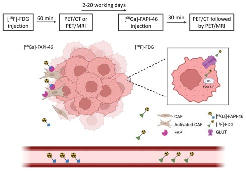
Introduction. [18F]FDG can be considered as PET tracer to determine whole-body staging in patients with locally advanced (LABC), metastatic, and recurrent breast cancer. Yet, previous studies demonstrated a lower amount of metabolic accumulation on [18F]FDG PET in case of oestrogen receptor (ER)-positive breast cancer. Consequently, given the lower metabolic accumulation, patients with ER-positive breast cancer might be at risk for underdiagnosis according to [18F]FDG PET. A novel PET tracer, [68Ga]-fibroblast activation protein inhibitor (FAPI)-46, has shown promising results in breast cancer (re)staging. This single-centre prospective pilot study aims to explore the relative diagnostic accuracy of [68Ga]-FAPI-46 compared to [18F]FDG in patients with ER-positive breast cancer and will provide the required information to support sample size calculation for a future validation study.
Methods. In this study, 20 patients with ER-positive LABC, metastatic, or recurrent breast cancer will undergo the standard [18F]FDG PET exam and a [68Ga]-FAPI-46 PET/CT and [68Ga]-FAPI-46 PET/MRI exam. Histopathological confirmation will be obtained in cases of discordant or concordant (double-positive) findings, depending on clinical relevance and technical feasibility. The study will provide preliminary estimates of the relative accuracy of [68Ga]-FAPI-46 PET when compared to [18F]FDG PET as well as the proportion of discordant findings and patients in whom a biopsy is feasible.
Conclusions. The present single-centre prospective pilot study will characterize the potential of [68Ga]-FAPI-46 PET to identify additional lesions not detected on a [18F]FDG PET exam in patients with ER-positive breast cancer to estimate an adequate sample size for a future validation study.
Trial registration. This study was registered prospectively on ClinicalTrials.gov. Trial registration number: NCT06335069; Register date: 28 March 2024.
Keywords: breast neoplasms, receptors, estrogen, FAPI-46, positron emission tomography computed tomography, discordant lesions
Introduction
Breast cancer is the most common type of invasive cancer among women, with more than 2.4 million new diagnoses worldwide in 2024 [1]. The prognosis of breast cancer patients is strongly influenced by the extent of the disease [2]. The five-year survival rate is 100% when confined to the breast, 87.2% with axillary lymph node involvement, and 32.6% with distant metastases [3]. Consequently, patients with more extensive disease at diagnosis, such as those with locally advanced breast cancer (LABC), require more comprehensive staging at diagnosis. LABC is typically defined as a primary tumour larger than 5 cm and/or the presence of lymph node metastases (cT3-4N0 or cT1-4N+) [4]. Patients with LABC, metastatic breast cancer, or recurrent breast cancer receive distant staging using [18F]-fluorodeoxyglucose ([18F]FDG) positron emission tomography (PET) [5]. [18F]FDG PET is combined with either computed tomography (CT) or magnetic resonance imaging (MRI) to obtain morphologic information [2].
Previous studies have shown that the amount of metabolism on [18F]FDG PET varies between breast cancer clinical subtypes, with a significantly lower metabolic activity in the oestrogen receptor (ER)-positive subtype [6-8]. Furthermore, Iqbal et al. assessed the diagnostic performance of [18F]FDG PET in a cohort of 70 ER-positive grade 1-2 breast cancer patients prior to treatment. Of the 155 lesions analysed, 7 (4.5%) were false negatives, affecting 7 patients, while 17 lesions (11%) were false positives in 9 patients. [18F]FDG PET staging of grade 1-2 ER-positive breast cancer resulted in misclassification in 22.9% of patients [8]. The lower metabolic activity in ER-positive, low-grade tumours can have important diagnostic consequences, as low FDG-avidity may lead to missed metastatic locations, potentially resulting in incorrect staging and treatment decisions [9-11].
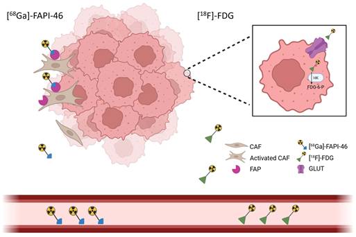
Given these limitations of [18F]FDG in ER-positive breast cancer, there is a need for an alternative PET tracer to improve baseline staging. A promising option is the fibroblast activation protein inhibitor (FAPI), which binds to fibroblast activation protein, a type II transmembrane serine protease expressed on the surface of activated cancer-associated fibroblasts within the tumour stroma (Figure 1). Fibroblast activation protein is highly expressed in more than 90% of epithelial-derived tumours and their metastases, whereas not expressed in healthy tissue, benign lesions, and precancerous tissues [12]. This characteristic makes it a potential target for both diagnosing and treating malignant tumours [12]. FAPI can be radiolabelled with Gallium-68 ([68Ga]) or with Fluorine-18 ([18F]), creating the 68Ga-FAPI or 18F-FAPI PET tracers. Previous studies with 68Ga-FAPI and 18F-FAPI have demonstrated enhanced lesion detectability, higher standardised uptake values (SUV) and improved tumour-to-background-ratios (TBR) across multiple cancer types, including breast cancer [13-17]. However, a direct comparison of 68Ga-FAPI PET and [18F]FDG PET in ER-positive breast cancer patients specifically has not yet been conducted.
Schematic illustration of the working principles of [68Ga]-FAPI-46 (left) and [18F]FDG (right) as PET tracers. [68Ga]-FAPI-46 binds to fibroblast activation protein (FAP), expressed by cancer-associated fibroblasts in the tumour microenvironment. Created with www.biorender.com.
Therefore, to address this gap, we propose a study design for a single-centre prospective pilot study to compare the relative diagnostic accuracy of [68Ga]-FAPI-46 and [18F]FDG as PET tracers in patients with ER-positive breast cancer.
Methods
Study design and patient selection
This single-centre prospective pilot study will compare the relative diagnostic accuracy of [68Ga]-FAPI-46-PET and [18F]FDG PET in patients with ER-positive breast cancer. All included patients will undergo [68Ga]-FAPI-46 PET/CT and [68Ga]-FAPI-46 PET/MRI in addition to their standard [18F]FDG PET exam. Histological results from biopsy will be used as reference standard. Biopsy will only be performed in patients who test positive on at least one tracer, verification of disease status in patients who test negative is not possible in the time frame of our study. This study will include female patients diagnosed with histopathological proven ER-positive LABC (cT3-4N0 or cT1-4N+), metastatic breast cancer, or recurrent breast cancer regardless of HER2 status who are scheduled to receive a whole-body [18F]FDG PET scan for distant staging (Table 1). Based on feasibility considerations, a total of 20 patients will be included, recruited at the Maastricht UMC+ (Maastricht, the Netherlands). Informed consent will be written, dated, and signed by the investigator and all participants. The study has been approved by the Medical Research Ethics Committee of Maastricht UMC+ via the clinical trial information system (CTIS; 2023-508066-15) and is registered at ClinicalTrials.gov (Identifier: NCT06335069). Monitoring will be conducted by the independent party Clinical Trial Centre Maastricht (CTCM).
Eligibility criteria
| Inclusion criteria |
| Female patient with histopathological proven ER-positive breast cancer (LABC (cT3-4N0 or cT1-4N+), metastatic, or recurrent breast cancer), regardless of HER2 status, at diagnosis |
| Distant staging by [18F]FDG PET/CT or [18F]FDG PET/MRI |
| Exclusion criteria |
| Age <18 |
| Pregnancy |
| Secondary malignancies |
| No [18F]FDG PET/CT or [18F]FDG PET/MRI exam |
| Contra-indication for PET/MRI (metallic device in body, severe claustrophobia, BMI > 35) |
| Chronic inflammatory disease (e.g. rheumatoid arthritis) |
| Severe hepatic or renal impairment (eGFR ≤ 45mL/min/1.73m²) as [68Ga]-FAPI-46 is primarily excreted through the kidneys and severe renal impairment may reduce tracer clearance and prolong circulation of the radiopharmaceutical. |
Radiopharmaceutical protocol
The precursor, FAPI-46 (SOFIE, Dulles, VA, USA), is labelled with the radioactive isotope [68Ga] at the radiopharmacy facility in Maastricht UMC+, following GMP-z compliant protocols and using an automated synthesis module (Scintomics, Germany). In short, [68Ga] is eluted from a [68Ge]/[68Ga] generator (Eckert-Ziegler) using 0.1 M HCl, adjusted to pH 3.5 using a HEPES buffer and combined with 50 microgram FAPI-46 precursor at 95 °C for 10 minutes. After synthesis, [68Ga]-FAPI-46 is purified using a Sep-PakC18 mini cartridge, where it is collected using 2 ml Ethanol/water (50/50) and diluted with 12 ml sterile PBS solution containing 0.1 mg ascorbic acid. Prior to injection, quality control of each radiosynthesis is performed, consisting of HPLC (identity, radiochemical and chemical purity), TLC (radiochemical purity), endotoxin test, pH and visual appearance.
Imaging protocol
All included patients will undergo [68Ga]-FAPI-46 PET/CT and [68Ga]-FAPI-46 PET/MRI in addition to their standard [18F]FDG PET exam. The time between the [18F]FDG PET exam and [68Ga]-FAPI-46 PET exam is required to be at least 48 hours to prevent potential interference from residual tracer activity. The maximum permitted time interval between scans will be 20 working days. To perform the [68Ga]-FAPI-46 PET/CT and PET/MRI scan, patients receive an intravenous injection of [68Ga]-FAPI-46 at a dose of 3 MBq/kg with a minimum of 100 MBq. The [68Ga]-FAPI-46 PET/CT scan will take place after a waiting period of 30 minutes, immediately followed by the [68Ga]-FAPI-46 PET/MRI scan [15, 18]. Neither exam involved the administration of a contrast agent. After acquisition, the [68Ga]-FAPI-46 PET scans will be evaluated for discordant lesions compared to the [18F]FDG PET scan. Based on this initial clinical assessment, a biopsy will be performed if clinically indicated and technically feasible, after discussion in the multidisciplinary team meeting. After completion of the study, two independent readers will re-evaluate all [18F]FDG PET and [68Ga]-FAPI-46 PET exams in a randomised order, assessing for parameters such as quantitative metabolic measurements and number of suspected lesions. This will be performed with a 8-week interval between [68Ga]-FAPI-46 and [18F]FDG assessments to minimize recall bias. Afterwards, both readers come together to reach consensus on their evaluations.
Histopathological biopsy
Histopathological biopsy will be considered for additional suspicious lesions detected during the PET exams, but not for the already histopathologically confirmed primary breast tumour. The decision for biopsy will be made on a lesion-by-lesion basis, depending on the tracers uptake pattern and the potential impact on clinical management. Specifically, biopsy will be considered in cases where the final interpretation indicates discordance between [68Ga]-FAPI-46 and [18F]FDG ([18F]FDG-positive and [68Ga]-FAPI-46-negative or [18F]FDG-negative and [68Ga]-FAPI-46-positive) or when an additional lesion is positive on both tracers but has not been previously confirmed. Whether a biopsy will be performed, will depend on two relevant conditions:
1. Does the additional lesion change the treatment choice? If the patient is already diagnosed with six or more hypermetabolic distant metastases according to [18F]FDG PET, no biopsy will be performed.
2. Technical feasibility of performing a biopsy. In some rare cases, depending on the anatomical location of the hypermetabolic lesion, it may be impossible to perform a biopsy. In such instances, these lesions will be mentioned for follow-up, with the duration of follow-up determined in consultation with clinicians.
Histopathological biopsy will be handled according to the standard of care.
Statistical analysis
This pilot study (n=20) will inform sample size estimation for a future validation study. As biopsy can only be performed in patients testing positive on at least one tracer, complete disease verification is not feasible. Longer follow-up will therefore be needed in subsequent studies. Nonetheless, 20 patients are sufficient to provide meaningful preliminary estimates [19]. Specifically, we will estimate: (1) the proportion of discordant pairs; (2) the proportion of lesions where biopsy is indicated and feasible; (3) the proportion of [68Ga]-FAPI-46-positive and/or [18F]FDG-positive lesions confirmed to have a histologically positive or negative lesion (gold standard); and (4) the proportion of lesions detected on [68Ga]-FAPI-46 PET/CT vs. [68Ga]-FAPI-46 PET/MRI, which will be calculated using the number of lesions detected by one specific modality divided by the total number of lesions detected by both modalities. The proportions (3) will be used for preliminary estimates of the relative true positive fraction and false positive fraction when comparing [68Ga]-FAPI-46 and [18F]FDG PET.
The analyses will be performed on a patient level. Statistical analysis will be performed using the Statistical Package for the Social Sciences (V28, IBM Corp, Armonk, New York, USA).
Discussion
Accurate staging of LABC (cT3-4N0 or cT1-4N+), metastatic-, and recurrent breast cancer, particularly in the ER-positive subtype, remains challenging due to the limitations of the current staging method by using [18F]FDG as PET tracer. ER-positive breast cancer lesions often exhibit lower metabolic activity on [18F]FDG PET compared to other breast cancer clinical subtypes, making this subgroup less suitable for staging using [18F]FDG PET as it may result in underestimation of the disease extent [7, 8, 20]. Given this limitation, alternative PET-tracers such as 68Ga-FAPI may provide a more effective solution, particularly for patients with ER-positive breast cancer. Consequently, we propose a single-centre prospective pilot study to compare the relative diagnostic accuracy of [68Ga]-FAPI-46 and [18F]FDG as PET tracers in patients diagnosed with ER-positive breast cancer.
FAPI is a relatively novel PET tracer in oncologic imaging. Therefore, in recent years, several studies have compared its diagnostic efficacy with [18F]FDG, the current standard of care [21, 22]. Multiple review articles across various cancer types revealed FAPI PET to offer superior sensitivity and specificity compared to [18F]FDG PET, with greater lesion detectability [23-26]. Moreover, [18F]FDG shows nonspecific uptake in crucial organs, whereas 68Ga-FAPI shows a drastic reduction of the background signal in the brain, liver, oro-and nasopharyngeal mucosa, and the gastrointestinal tract, resulting in a higher TBR [27]. In addition, previous studies concerning 68Ga-FAPI in breast cancer, not solely focusing on the ER-positive breast cancer subtype, have reported a higher detection rate and higher SUV values for 68Ga-FAPI PET compared to [18F]FDG PET within the breast, axillary lymph nodes, and distant metastases [15-17].
Despite the promising preliminary evidence, no prospective study has specifically compared FAPI PET with [18F]FDG PET in ER-positive breast cancer. To address this gap, this study is designed to provide insights on this topic. The primary aim of this pilot study is to obtain data on the relative performance of both tracers and the feasibility of taking biopsies in test-positive patients. If FAPI PET detects more lesions compared to [18F]FDG PET, it may be considered superior, however, it would not be preferable if this comes at the cost of a higher rate of false-positive results. The results of this study will be valuable for assessing the feasibility of a larger, potentially multicentre study, and identifying potential challenges in the study workflow. While this pilot study is not intended to detect subtle statistical differences, it will generate insights into essential input parameters for an adequate sample size calculation for a diagnostic study that compares the diagnostic accuracy of [18F]FDG PET and [68Ga]-FAPI-46 as PET tracers in patients with ER-positive breast cancer.
A major strength of the study design is its prospective approach, which allows for systematic data collection and minimises the risk of bias. The use of standardised imaging protocols increases reproducibility and ensures consistent acquisition across patients. Furthermore, the blinded image interpretation limits observer-related variability and strengthens the reliability of the results. Finally, the single-centre setting will facilitate strict protocol adherence, supporting strong internal validation.
Despite these strengths, several limitations should be acknowledged. An important limitation of 68Ga-FAPI is the potential of false positive findings, since non-malignant fibrotic processes and chronic inflammatory diseases such as fibroadenoma, liver fibrosis, and rheumatoid arthritis are associated with fibroblast activity, and may exhibit 68Ga-FAPI uptake [17, 28]. However, this potential limitation is partly mitigated by the integration of multimodal imaging in this study ([18F]FDG PET, CT, and MRI), which provides an opportunity to further characterize such lesions. By correlating these findings with histopathology when available, the imaging data may help to distinguish benign from malignant uptake patterns. This approach could reduce the need for unnecessary biopsies, as lesions suspected to represent false positives may be identified through imaging characteristics alone. Although multimodal imaging is an appropriate strategy for the current study, the independent use of 68Ga-FAPI PET as a stand-alone imaging modality could be more limited, given the reduced ability to correlate uptake with anatomical or other functional imaging findings.
Nonetheless, a key remaining limitation of this study is that when a lesion appears negative on the 68Ga-FAPI scan, no biopsy will be performed. As a result, false-negative and true-negative findings cannot be estimated, and sensitivity, specificity, NPV, and accuracy remain unknown. Only the relative accuracy of 68Ga-FAPI and [18F]FDG can be estimated [19]. Future research may provide further insights by incorporating longer follow-up.
In addition to comparing the relative diagnostic accuracy of [68Ga]-FAPI-46 with [18F]FDG, this study will provide essential feasibility data, optimise scanning protocols, and identify potential logistical or methodological obstacles, laying the groundwork for a diagnostic study. In particular, by performing 68Ga-FAPI PET/CT immediately followed by 68Ga-FAPI PET/MRI with a single tracer injection, the study will generate insights not only into the imaging parameters, but also into the practical workflow, timing, and coordination required to acquire high-quality images across two modalities. Eventually, these results will guide future imaging strategies in ER-positive breast cancer, potentially impacting lesion detection, staging accuracy, and treatment planning. Based on the results of this pilot study, including the relative accuracy and available histopathology, it will be possible to estimate the number of patients required for a future validation study to robustly assess sensitivity, specificity, and the feasibility of taking biopsies.
Conclusion
[68Ga]-FAPI-46 is a highly promising novel PET tracer, though it has not yet been investigated specifically in patients with ER-positive breast cancer. This single-centre prospective pilot study will compare the relative diagnostic accuracy of [68Ga]-FAPI-46 and [18F]FDG as PET tracer in patients with ER-positive LABC, metastatic, and recurrent breast cancer.
Abbreviations
[18F]FDG: [18F]-fluorodeoxyglucose; [68Ga]: Gallium-68; CT: Computed tomography; CTCM: Clinical trial Centre Maastricht; CTIS: Clinical trial information system; ER: Oestrogen receptor; FAPI: Fibroblast activation protein inhibitor; GMP: Good manufacturing practice; HER2: Human epidermal growth factor receptor 2; LABC: Locally advanced breast cancer; MRI: Magnetic resonance imaging; PET: Positron emission tomography; SUV: Standardised uptake value; TBR: Tumour-to-background ratio.
Acknowledgements
Funding
M. Lenaerts received salary from Kankeronderzoeksfonds Limburg. L. Duijx received salary from the Academic Fund of Maastricht UMC+.
Author contributions
TN conceived the original idea and proposed the study concepts. ML and LD prepared the manuscript. ML, TN, MB, and RG were involved in the ethical approval of the study. PN verified the statistical analysis. ML, LD, FM, MB, RG, VTH, PB, LK, PN, MS, JW and TN were responsible for the manuscript review. All authors have read and approved the manuscript.
Ethics approval and consent to participate
The study has been approved by the Medical Research Ethics Committee of Maastricht UMC+ via the clinical trial information system (CTIS; 2023-508066-15). Informed consent will be written, dated, and signed by the investigator and all participants.
Competing Interests
F.M.M. is supported by the German Research Foundation within the framework of Research Training Group 2375 (Tumour-Targeted Drug Delivery; Grant 331065168) and Clinical Research Unit 5011 (“Integrating Emerging Methods to Advance Translational Kidney Research [InteraKD]”; Project 445703531) and reports institutional grants/contracts from GE Precision Healthcare LLC, NanoMab Technology Ltd., Radiopharm Ltd., and Siemens Healthcare not related to the study; and personal/consulting fees from Advanced Accelerator Applications (AAA) GmbH/Novartis, CURIUMTM, NanoMab Technology Ltd., and Telix Pharmaceuticals outside the submitted work. V.C.G.T.H. reports institutional grants and personal fees from Roche, Novartis, Pfizer, Eli Lilly, and institutional grants from AstraZeneca, Daiichi Sankyo, and Gilead. P.B. received speaker honoraria from GE Healthcare and Siemens, not related to the content of this study. L.F.S.K. received speaker honorarium for education from SCEM, paid to the hospital, not related to the content of this study. M.L.S. received institutional research funding not related to this study from Servier Pharma, Nutricia and Illumina for microbiota research & Roche for unrelated research. J.E.W. reports institutional support from Abbott, Anaconda Biomed, Asklepios, Bayer, Becton & Dickinson Medical, Bentley, Boston, Brainlab, GE Healthcare, Gleamer, Hologic, Inari Medical, Johnson & Johnson, LCRB, Medtronic, Merit Medical Systems, Microvention, Nico-Lab, Nova Techs, Oldelft Benelux, Ontario Association of Radiologists, Penumbra, Philips, Screenpoint Medical, Siemens, Stryker, Tajpan Sro and reports speaker honoraria from Bayer and Siemens not related to content of this study. T.J.A.v.N received speaker honoraria, participation of medical advisory boards and institutional grant support from Bayer and GE Healthcare, not related to the content of this study and reports consultancy support for Screenpoint Medical, not related to the content of this study. All other authors declare to have no conflicts of interest.
References
1. Sung H, Filho AM, Laversanne M, Ferlay J, Siegel RL, Soerjomataram I. et al. Global cancer statistics 2024: GLOBOCAN estimates of incidence and mortality worldwide for 34 cancers in 186 countries. CA Cancer J Clin. 2026;76:e70090
2. Gennari A, Andre F, Barrios CH, Cortes J, de Azambuja E, DeMichele A. et al. ESMO Clinical Practice Guideline for the diagnosis, staging and treatment of patients with metastatic breast cancer. Ann Oncol. 2021;32:1475-95
3. SEER. Cancer Stat Facts: Female Breast Cancer. 2021.
4. Simos D, Clemons M, Ginsburg OM, Jacobs C. Definition and consequences of locally advanced breast cancer. Curr Opin Support Palliat Care. 2014;8:33-8
5. Groheux D. FDG-PET/CT for Primary Staging and Detection of Recurrence of Breast Cancer. Semin Nucl Med. 2022;52:508-19
6. Avril S, Muzic RF Jr, Plecha D, Traughber BJ, Vinayak S, Avril N. (1)(8)F-FDG PET/CT for Monitoring of Treatment Response in Breast Cancer. J Nucl Med. 2016;57(Suppl 1):34S-9S
7. de Mooij CM, Ploumen RAW, Nelemans PJ, Mottaghy FM, Smidt ML, van Nijnatten TJA. The influence of receptor expression and clinical subtypes on baseline [18F]FDG uptake in breast cancer: systematic review and meta-analysis. EJNMMI Res. 2023;13:5
8. Iqbal R, Mammatas LH, Aras T, Vogel WV, van de Brug T, Oprea-Lager DE. et al. Diagnostic Performance of [(18)F]FDG PET in Staging Grade 1-2, Estrogen Receptor Positive Breast Cancer. Diagnostics (Basel). 2021 11
9. Schäfer L, Altunay B, Heesch A, van Nijnatten T, Vaz S, Juweid ME. et al. Current Approaches of Nuclear Molecular Imaging in Breast Cancer. Cancers. 2025;17:2105
10. Jalaguier-Coudray A, Delarbre B, Brenot-Rossi I, Houvenaeghel G, Villard-Mahjoub R, Viens P. et al. Contribution of FDG PET/CT for the Optimization of the Management of Additional Lesions Detected on Local Staging Breast MRI. AJR Am J Roentgenol. 2016;206:891-900
11. Abouzied MM, Crawford ES, Nabi HA. 18F-FDG imaging: pitfalls and artifacts. J Nucl Med Technol. 2005;33:145-55 quiz 62-3
12. Huang R, Pu Y, Huang S, Yang C, Yang F, Pu Y. et al. FAPI-PET/CT in Cancer Imaging: A Potential Novel Molecule of the Century. Front Oncol. 2022;12:854658
13. Mona CE, Benz MR, Hikmat F, Grogan TR, Lueckerath K, Razmaria A. et al. Correlation of (68)Ga-FAPi-46 PET Biodistribution with FAP Expression by Immunohistochemistry in Patients with Solid Cancers: Interim Analysis of a Prospective Translational Exploratory Study. J Nucl Med. 2022;63:1021-6
14. Kratochwil C, Flechsig P, Lindner T, Abderrahim L, Altmann A, Mier W. et al. (68)Ga-FAPI PET/CT: Tracer Uptake in 28 Different Kinds of Cancer. J Nucl Med. 2019;60:801-5
15. Zheng S, Lin J, Zhu Y, Chen Y, Zhang J, Chen X. et al. 68Ga-FAPI Versus 18F-FDG PET/CT in Evaluating Newly Diagnosed Breast Cancer Patients: A Head-to-Head Comparative Study. Clin Nucl Med. 2023;48:e104-e9
16. Komek H, Can C, Guzel Y, Oruc Z, Gundogan C, Yildirim OA. et al. (68)Ga-FAPI-04 PET/CT, a new step in breast cancer imaging: a comparative pilot study with the (18)F-FDG PET/CT. Ann Nucl Med. 2021;35:744-52
17. Elboga U, Sahin E, Kus T, Cayirli YB, Aktas G, Uzun E. et al. Superiority of (68)Ga-FAPI PET/CT scan in detecting additional lesions compared to (18)FDG PET/CT scan in breast cancer. Ann Nucl Med. 2021;35:1321-31
18. Backhaus P, Burg MC, Asmus I, Pixberg M, Buther F, Breyholz HJ. et al. Initial Results of (68)Ga-FAPI-46 PET/MRI to Assess Response to Neoadjuvant Chemotherapy in Breast Cancer. J Nucl Med. 2023;64:717-23
19. Alonzo TA, Kittelson JM. A novel design for estimating relative accuracy of screening tests when complete disease verification is not feasible. Biometrics. 2006;62:605-12
20. Bin J, Wenjia L, Wan W, Kaiyin M, Bowen Y, Qichang W. et al. Comparison of (18)F-FDG PET/CT and (18)F-FAPI PET/CT in Systemic Staging of Newly Diagnosed Breast Cancer. Acad Radiol. 2024;32:50-7
21. Guo W, Pang Y, Yao L, Zhao L, Fan C, Ke J. et al. Imaging fibroblast activation protein in liver cancer: a single-center post hoc retrospective analysis to compare [(68)Ga]Ga-FAPI-04 PET/CT versus MRI and [(18)F]-FDG PET/CT. Eur J Nucl Med Mol Imaging. 2021;48:1604-17
22. Evangelista L, Filippi L, Schillaci O. What radiolabeled FAPI pet can add in breast cancer? A systematic review from literature. Ann Nucl Med. 2023;37:442-50
23. Abbasi S, Dehghani M, Khademi S, Irajirad R, Parizi ZP, Sahebi M. et al. Revolutionizing cancer diagnosis and dose biodistribution: a meta-analysis of [68ga] FAPI- 46 vs. [18f] FDG imaging. Syst Rev. 2025;14:109
24. Treglia G, Muoio B, Roustaei H, Kiamanesh Z, Aryana K, Sadeghi R. Head-to-Head Comparison of Fibroblast Activation Protein Inhibitors (FAPI) Radiotracers versus [(18)F]F-FDG in Oncology: A Systematic Review. Int J Mol Sci. 2021 22
25. Ruan D, Zhao L, Cai J, Xu W, Sun L, Li J. et al. Evaluation of FAPI PET imaging in gastric cancer: a systematic review and meta-analysis. Theranostics. 2023;13:4694-710
26. Duijx L, Lenaerts M, Smidt ML, Vaz SC, Backhaus P, Bauwens M. et al. Diagnostic Performance of FAPI PET in Patients with Breast Cancer: A Systematic Review. J Nucl Med. 2026
27. Mori Y, Dendl K, Cardinale J, Kratochwil C, Giesel FL, Haberkorn U. FAPI PET: Fibroblast Activation Protein Inhibitor Use in Oncologic and Nononcologic Disease. Radiology. 2023;306:e220749
28. Radmehr K, Farzanefar S, Abbasi M, Salehi Y, Karamzade-Ziarati N, Emami-Ardekani A. et al. Comparison of diagnostic performance of [(68)Ga]-Ga-FAPI-46 and [(18)F]-FDG PET/CT imaging for the detection of lesions and disease staging in patients with breast cancer. Asia Ocean J Nucl Med Biol. 2025;13:1-9
Author contact
Corresponding author: Melissa Lenaerts, Department of Surgery, Maastricht University Medical Centre+, P.O. Box 5800, 6202 AZ Maastricht, The Netherlands. ORCID: 0000-0001-9687-4375. melissa.lenaertsnl.