Impact Factor ISSN: 1837-9664




J Cancer 2026; 17(9):1615-1625. doi:10.7150/jca.138124 This issue Cite
Research Paper
Chronically Established ρ0 Cancer Cells Exhibit Shared Stemness-Associated and Aggressive Phenotypes Across Multiple Cancer Cell Lines
Yu-Seon Han1*, Myeong-Eun Jegal3*, Su Yeon Lee1, Joung-Sun Park1, Yung-Jin Kim1,2, 
1. Institute of Nanobio Convergence, Pusan National University, Busan 46241, Republic of Korea.
2. Department of Molecular Biology, Pusan National University, Busan 46241, Republic of Korea.
3. Marine Biotechnology & Bioresource Research Department, Korea Institute of Ocean Science & Technology, Busan, Republic of Korea.
*These authors contributed equally to this work.
Received 2026-5-21; Accepted 2026-8-12; Published 2026-9-3
Abstract
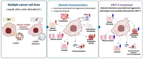
Chronically established mitochondrial DNA-depleted (ρ0) cancer cells may exhibit phenotypic changes beyond metabolic dysfunction; however, whether such changes are shared across different cancer types remains unclear. In this study, we generated chronically established ρ0 models from pancreatic, lung, and breast cancer cell lines and examined their common phenotypic features. Compared with their parental wild-type counterparts, the ρ0 cell lines exhibited increased proliferation, chemoresistance-related features, sphere-forming ability, wound closure, invasion-related phenotypes, and angiogenic activity, together with common morphological alterations. These changes were accompanied by VDAC1 upregulation and cytoskeletal remodeling. Pharmacologic treatment with VBIT-4 attenuated several of these phenotypes, whereas its effects on chemoresistance-related responses varied across cell lines. Collectively, these findings suggest that chronically established ρ0 cancer cells exhibit shared stemness-associated and aggressive phenotypes across multiple cancer cell lines, accompanied by VDAC1 upregulation and cytoskeletal remodeling.
Keywords: mtDNA depletion, ρ0 cells, cancer cell plasticity, stemness-associated phenotype, cytoskeletal remodeling
Introduction
Mitochondria play essential roles in cellular energy production, biosynthesis, apoptosis, redox regulation, and intracellular signaling [1, 2]. Mitochondrial dysfunction has been implicated in a wide range of diseases and is increasingly recognized as an important feature of tumor progression and cellular adaptation in cancer [2-7]. However, the extent to which chronic mitochondrial DNA (mtDNA) depletion is associated with shared phenotypic changes across different cancer types remains incompletely understood.
We previously demonstrated that mtDNA depletion-associated mitochondrial dysfunction in Hep3B cells was associated with stemness-associated and aggressive cancer cell phenotypes [8]. Mitochondrial pathways have also been implicated in cancer cell plasticity and stemness-associated phenotypes [9]. Wnt signaling is likewise widely associated with stemness-associated phenotypes and cancer cell plasticity [10-12]. Cancer cells exhibiting stemness-associated phenotypes can display properties such as sphere-forming ability, chemoresistance, wound closure and invasion-related behavior, and pro-angiogenic activity, all of which are often associated with tumor progression, recurrence, and metastasis [10-12]. Whether these phenotypic changes are shared among chronically established mtDNA-depleted cancer cells derived from different tissue origins remains unclear.
Voltage-dependent anion channels (VDACs) are mitochondrial porins located on the outer mitochondrial membrane that participate in metabolite and ion transport, reactive oxygen species release, apoptotic signaling, and regulation of mitochondrial membrane permeability [13-16]. The cytoskeleton, comprising microfilaments, microtubules, and intermediate filaments, maintains cell shape and structural integrity and is closely associated with mitochondrial dynamics and organelle organization [16, 17]. These functional relationships suggest that alterations in mitochondrial membrane-associated factors and cytoskeletal organization may be relevant to the phenotypic changes observed in chronically established ρ0 cells.
In this study, we examined whether chronically established ρ0 cells derived from pancreatic, lung, and breast cancer cell lines exhibit shared stemness-associated and aggressive phenotypes. We further investigated whether these changes are accompanied by VDAC1 upregulation and cytoskeletal remodeling and whether pharmacologic treatment with VBIT-4 alters selected phenotypes observed in ρ0 cells.
Materials and Methods
Materials
Dulbecco's modified Eagle's medium (DMEM), RPMI 1640, and fetal bovine serum (FBS) were purchased from JBI (Daegu, Republic of Korea). EGM®-2 BulletKit®, EGM® BulletKit®, and ReagentPack™ subculture reagents were obtained from Lonza (Basel, Switzerland). TRIzol reagent was purchased from QIAGEN (Venlo, Netherlands). Reverse Transcriptase Premix and PCR Premix were purchased from Elpis Biotech and Bioneer (Daejeon, Republic of Korea), respectively. Pro-PREP™ protein extraction solution was obtained from iNtRON Biotechnology (Daejeon, Republic of Korea). Primary antibodies were purchased from Cell Signaling Technology (Danvers, MA, USA), Proteintech (Rosemont, IL, USA), and Santa Cruz Biotechnology (Santa Cruz, CA, USA). Alexa Fluor™ 488, DAPI, MitoTracker™ Red CMXRos, and phalloidin were purchased from Invitrogen (Carlsbad, CA, USA). Doxorubicin (DX), ethidium bromide, rotenone, and antimycin A were purchased from Sigma-Aldrich (St. Louis, MO, USA). VBIT-4 was purchased from Aobious (Gloucester, MA, USA). The In Situ Cell Death Detection Kit was purchased from Roche (Indianapolis, IN, USA). Matrigel and Transwell inserts with 8-μm pores were purchased from Corning (Corning, NY, USA).
Cell culture
Hep3B and MDA-MB-231 cells were obtained from the Korean Cell Line Bank (Seoul, Republic of Korea), and HPAC and A549 cells were obtained from the American Type Culture Collection (Manassas, VA, USA). HPAC, A549, and MDA-MB-231 cells were maintained in RPMI 1640 supplemented with 10% FBS and 25 μg/mL amphotericin B at 37℃ in a humidified atmosphere containing 5% CO2. Hep3B cells were maintained in DMEM supplemented with 10% FBS and 25 μg/mL amphotericin B under the same conditions. Hep3B/ρ0 cells had been established previously and were maintained in our laboratory, whereas HPAC/ρ0, A549/ρ0, and MDA-MB-231/ρ0 cells were newly established in this study. All ρ0 cells were maintained in DMEM supplemented with 10% FBS, 50 μg/mL uridine, 1 mM sodium pyruvate, and 25 μg/mL amphotericin B at 37℃ in 5% CO2.
Human umbilical vein endothelial cell (HUVEC) culture
HUVECs (Lonza) were maintained in EGM™-2 medium supplemented with the EGM™-2 Endothelial SingleQuots™ kit at 37℃ in 5% CO2. Cells at passages 4-7 were used for experiments.
Establishment of ρ0 cell lines
HPAC/ρ0, A549/ρ0, and MDA-MB-231/ρ0 cells were established by long-term treatment with ethidium bromide. Parental cells were cultured for 2-3 weeks in complete DMEM containing 1 μg/mL ethidium bromide, after which candidate ρ0 cells were selected by treatment with rotenone and antimycin A. After selection and confirmation of mtDNA depletion, the cells were maintained in ethidium bromide-free ρ0 maintenance medium. Loss of mitochondrial gene expression was used as an indicator of mtDNA depletion.
Reverse transcriptase-polymerase chain reaction (RT-PCR)
Total RNA was isolated using TRIzol reagent according to the manufacturer's instructions. Complementary DNA was synthesized from 2 μg of total RNA using Reverse Transcriptase Premix. PCR amplification was performed in a total volume of 20 μL containing PCR Premix and gene-specific primers. The amplification conditions were as follows: 30 cycles of denaturation at 95℃ for 20 s, annealing at 57℃ for 10 s, and extension at 72℃ for 40 s. PCR products were separated by electrophoresis on 1% agarose gels, stained with GelRed™, and visualized under UV illumination. The expression of mtDNA-encoded genes (COX1, COX2, COX3, ND1, ND2, CYTB, and ATP6) was examined to assess depletion of mitochondrial transcripts in ρ0 cells. Primer sequences are provided in Table 1.
Primer sequences used for RT-PCR
| Gene name | Sequence |
|---|---|
| COX1 | 5ʹ-TTG AAC AGT CTA CCC TCC C-3ʹ |
| 5ʹ-GCT CAC ACG ATA AAC CCT-3ʹ | |
| COX2 | 5ʹ-CGT CTG AAC TAT CCT GCC-3ʹ |
| 5ʹ-GTC GTG TAG CGG TGA AAG-3ʹ | |
| COX3 | 5ʹ-GAA AGC ACA TAC CAA GGC-3ʹ |
| 5ʹ-GCG GAT GAA GCA GAT AGT-3ʹ | |
| ND1 | 5ʹ-GAG CAG TAG CCC AAA CAA-3ʹ |
| 5ʹ-TAG GGT GAG TGG TAG GAA GT-3ʹ | |
| ND2 | 5ʹ-ACC CGT CAT CTA CTC TAC CA-3ʹ |
| 5ʹ-TAA TCC ACC TCA ACT GCC-3ʹ | |
| CYTB | 5ʹ-CAC TCC ACC TCC TAT TCT TG-3ʹ |
| 5ʹ-CTT ACT GGT TGT CCT CCG AT-3ʹ | |
| ATP6 | 5ʹ-GTT CGC TTC ATT CAT TGC-3ʹ |
| 5ʹ-TGA GTA GGC TGA TGG TTT C-3ʹ | |
| GAPDH | 5ʹ-ATC TTC CAG GAG CGA GAT CCC-3ʹ |
| 5ʹ-AGT GAG CTT CCC GTT CAG CTC-3ʹ |
Crystal violet assay
For proliferation assays, cells were seeded at 2 × 10⁴ cells/well in 24-well plates and incubated for 48 h in their respective maintenance media. For drug response assays, cells were incubated overnight and subsequently treated with 2 μM DX and/or 10 μM VBIT-4 for 48 h. Following treatment, the cells were stained with 1 mg/mL crystal violet for 10 min, washed, and dissolved in 10% SDS. Absorbance was measured at 570 nm.
TUNEL assay
Apoptotic cell death was assessed using the In Situ Cell Death Detection Kit (Roche). Cells were incubated overnight and subsequently treated with 2 μM doxorubicin for 48 h. The cells were fixed with paraformaldehyde, permeabilized, and incubated with the TUNEL reaction mixture for 1 h at 37 °C in the dark. Fluorescence images were acquired using an iRiS™ Digital Cell Imaging System (Logos Biosystems, Gyeonggi-do, Republic of Korea) at 200× magnification.
Western blotting
Total cell lysates were prepared using Pro-PREP™ extraction buffer supplemented with 1 mM sodium orthovanadate. Equal amounts of protein (30 μg) were separated by electrophoresis on 10% SDS-polyacrylamide gels and transferred onto polyvinylidene difluoride membranes. Membranes were incubated with appropriate primary antibodies diluted 1:1000, followed by incubation with horseradish peroxidase-conjugated secondary antibodies. Protein signals were detected using enhanced chemiluminescence reagents.
Wound-healing assay
Cells (1 × 10⁶) were seeded in 60-mm dishes and incubated overnight. A linear wound was generated using a razor blade, after which cells were maintained for up to 24 h in medium containing 1% FBS with or without 10 μM VBIT-4. RPMI 1640 was used for HPAC, A549, and MDA-MB-231 cells, whereas DMEM was used for Hep3B and all ρ0 cells. Wound closure was assessed by acquiring images of wounded areas at 40× magnification between 16 and 24 h after wounding, and cells that migrated beyond the reference line were counted.
Transwell invasion assay
Cell invasion was assessed using 24-well Transwell chambers fitted with 8-μm-pore membranes coated with Matrigel (1 mg/mL). Cells (2 × 10⁴) were suspended in medium containing 1% FBS with or without 10 μM VBIT-4 and seeded into the upper chambers. The lower chambers contained the corresponding maintenance medium for each cell line: RPMI 1640 for HPAC, A549, and MDA-MB-231 cells, and DMEM for Hep3B and all ρ0 cells. After 18 h of incubation, cells that had invaded to the lower surface of the membrane were fixed, stained, and counted under a microscope at 100× magnification.
Conditioned medium (CM) preparation
Cells (1 × 10⁶) were seeded in 100-mm dishes in their respective maintenance media and incubated overnight. After washing with PBS, the medium was replaced with medium containing 1% FBS: RPMI 1640 for HPAC, A549, and MDA-MB-231 cells, and DMEM for Hep3B and all ρ0 cells. After 24 h, the CM was collected, centrifuged to remove debris, and filtered through a 0.22-μm filter. CM was freshly prepared for experiments or stored at -80 °C until use.
Tube formation assay
A 96-well plate was coated with Matrigel at 60 μL/well and allowed to polymerize. HUVECs (2 × 10⁴ cells/well) were seeded onto the polymerized Matrigel layer and incubated in a mixture of EGM and CM at a 7:3 ratio. CM derived from wild-type (WT) or ρ0 cells was used as indicated, and 10 μM VBIT-4 was added where applicable. After 6 h of incubation, tube-like structures were imaged at 100× magnification. Quantitative analysis was performed using the Angiogenesis Analyzer plugin in ImageJ.
Immunofluorescence staining
Cells (2 × 10⁴) were cultured on glass coverslips for 24 h with or without 10 μM VBIT-4. For mitochondrial staining, the cells were incubated with 200 nM MitoTracker for 30 min. The cells were then fixed with 3.7% formaldehyde and permeabilized with 0.1% Triton X-100. The cells were incubated with primary antibodies against VDAC1, β-tubulin, or vimentin, followed by incubation with Alexa Fluor™ 488-conjugated secondary antibodies. F-actin was visualized using phalloidin, and nuclei were counterstained with DAPI. Images were acquired using an iRiS™ Digital Cell Imaging System (Logos Biosystems) at 200× magnification.
Sphere formation assay
Sphere-forming ability was assessed using non-adherent 96-well plates precoated with 0.4% agarose. One cell was seeded per well to minimize aggregation-derived sphere formation and cultured for 14 days in their respective maintenance media. Sphere formation was assessed based on the presence or absence of sphere formation in each well at the end of the culture period. For VBIT-4 treatment experiments, cells were exposed to 10 μM VBIT-4 throughout the culture period.
Protein-protein interaction network analysis
Protein-protein interaction network analysis was performed using the STRING database. Selected mitochondrial, cytoskeletal, membrane-associated, and stemness-related factors were used as input based on the phenotypic and molecular changes observed in ρ0 cells. The resulting network was organized to better facilitate visualization of the relationships involving VDAC family proteins.
Statistical analysis
Data are presented as mean ± standard deviation (SD). Statistical analysis was performed using GraphPad Prism software (version 9; GraphPad, San Diego, CA, USA). Two-group comparisons were performed using Student's t-test, whereas multiple-group comparisons were performed using one-way analysis of variance (ANOVA) followed by Tukey's multiple comparison test. Analyses involving two independent variables were performed using two-way ANOVA. Statistical significance was set at P < 0.05.
Results
Chronically established ρ0 cells exhibit common structural and growth-related changes
To investigate whether chronically established mtDNA-depleted (ρ0) cells exhibit common phenotypic changes across different cancer types, we generated ρ0 cell lines from pancreatic, lung, and breast cancer cell lines. Because mtDNA depletion is a defining characteristic of ρ0 cells, the expression of mtDNA-encoded genes, namely COX1, COX2, COX3, ND1, ND2, CYTB, and ATP6, was examined. All established ρ0 cell lines exhibited complete loss of expression of these mitochondrial genes (Figure 1A). In addition, MitoTracker staining revealed markedly reduced mitochondrial content in ρ0 cells compared with their parental WT counterparts (Figure 1B), confirming successful establishment of the mtDNA-depleted ρ0 models.
Establishment and common features of chronically established ρ0 cells. (A) RT-PCR was performed to examine mitochondrial gene expression in wild-type (WT) and ρ0 cells, demonstrating loss of mitochondrial gene expression in ρ0 cells. (B) MitoTracker staining was performed to compare mitochondrial content between WT and ρ0 cells and revealed a marked reduction in ρ0 cells. (C) Cell proliferation was assessed by crystal violet assay, demonstrating increased growth of ρ0 cells compared with WT cells. (D) Bright-field images show morphological differences between WT and ρ0 cells. The red boxes highlight the regions shown at higher magnification in the panels below. Data are presented as mean ± SD. ****P < 0.0001; ***P < 0.001; **P < 0.01; *P < 0.05; ns, not significant. 231 denotes MDA-MB-231.
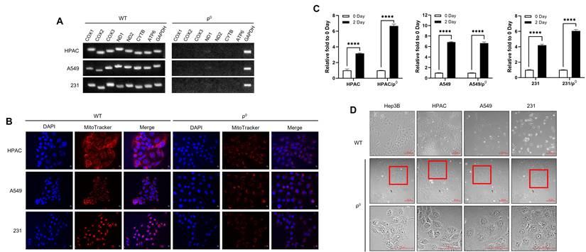
Despite their different tissue origins, the ρ0 cell lines exhibited several common features. All ρ0 cell lines showed increased proliferation rates compared with WT cells, with increases of approximately 1.3- to 2-fold depending on the cell line (Figure 1C). The greatest increase was observed in the HPAC-derived ρ0 cells. Bright-field microscopy further revealed that WT cells exhibited distinct morphologies depending on their tissue origins, whereas ρ0 cells consistently exhibited a rounded and flattened morphology (Figure 1D). Collectively, these findings demonstrate that chronically established ρ0 cells exhibit common structural and growth-related changes across multiple cancer cell lines of different tissue origins.
Chronically established ρ0 cells exhibit stemness-associated and aggressive phenotypes
We next examined whether chronically established ρ0 cells exhibit enhanced sphere-forming ability under single-cell culture conditions designed to minimize aggregation-derived sphere formation. None of the WT cell lines formed spheres, whereas all ρ0 cell lines formed spheres from single cells (Figure 2A). Although sphere morphology and size varied among the ρ0 cell lines, sphere-forming efficiency was consistently higher in ρ0 cells than in WT cells. To further characterize molecular changes associated with the ρ0 state, we analyzed the expression of Wnt3 and Wnt5. Most ρ0 cell lines exhibited decreased Wnt3 expression and increased Wnt5 expression compared with WT cells; however, HPAC/ρ0 cells exhibited increased Wnt3 expression compared with HPAC WT cells (Figure 2B). These findings indicate that alterations in Wnt-related signaling components associated with the ρ0 state vary among cell lines.
Stemness-associated and aggressive features in chronically established ρ0 cells. (A) Sphere formation was assessed under single-cell-per-well culture conditions and was observed in ρ0 cells but not in WT cells. The number of sphere-forming wells was quantified as a measure of sphere-forming ability. (B) Wnt3 expression was decreased and Wnt5 expression was increased in most ρ0 cell lines, whereas HPAC/ρ0 cells exhibited increased Wnt3 expression compared with WT cells. (C) Chemoresistance was assessed after treatment with doxorubicin (DX), and ρ0 cells exhibited greater resistance than WT cells. (D) TUNEL staining was performed to detect apoptotic cells following DX treatment. Apoptotic signals were observed in WT cells but not in ρ0 cells. (E) Following DX treatment, cleaved caspase-3 was detected in WT cells but not in ρ0 cells. The PARP antibody used recognizes both full-length and cleaved PARP; under these conditions, PARP cleavage was detected in WT cells, whereas no detectable cleaved PARP band was observed in ρ0 cells. ABCB1 expression was increased in ρ0 cells. (F) Wound closure was assessed using a wound-healing assay and quantified, demonstrating enhanced wound closure in ρ0 cells under reduced-serum conditions. (G) Cell invasion was assessed using a Transwell assay, and the number of invading cells was quantified, demonstrating the increased invasive ability of ρ0 cells. (H) In most ρ0 cell lines, E-cadherin expression was decreased, whereas N-cadherin and vimentin expression were increased; however, vimentin expression was decreased in MDA-MB-231/ρ0 cells. (I) Tube formation was assessed in human umbilical vein endothelial cells (HUVECs) exposed to WT- and ρ0-derived conditioned media. Quantitative ImageJ-based analysis demonstrated increased angiogenic activity in the ρ0 conditioned media group. (J) Expression of the angiogenic factors TGF-β and VEGF was increased in ρ0 cells. Data are presented as mean ± SD. ****P < 0.0001; ***P < 0.001; **P < 0.01; *P < 0.05; ns, not significant. 231 denotes MDA-MB-231.
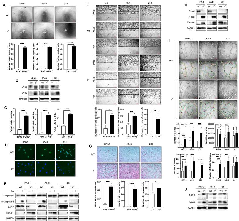
We next determined whether chronically established ρ0 cells exhibit altered responses to doxorubicin treatment, including chemoresistance-related features. Following doxorubicin treatment, WT cells exhibited a marked reduction in cell number, whereas ρ0 cells primarily exhibited reduced cell growth rather than overt cell death (Figure 2C). Consistent with this, TUNEL assays revealed TUNEL-positive cells in WT cells but not in ρ0 cells following doxorubicin treatment (Figure 2D). Western blot analysis further supported these findings, as cleaved caspase-3 was detected in WT cells but not in ρ0 cells. The PARP antibody used in this study recognizes both full-length and cleaved PARP; under our experimental conditions, PARP cleavage was detected in WT cells following doxorubicin treatment, whereas no detectable cleaved PARP band was observed in ρ0 cells. In addition, ABCB1 expression was higher in ρ0 cells under basal conditions and further increased following doxorubicin treatment (Figure 2E). Collectively, these results indicate that the chronically established ρ0 state is associated with enhanced chemoresistance-related features and reduced apoptotic responses to doxorubicin.
We next examined whether wound closure and invasion-related phenotypes were altered in ρ0 cells. Wound-healing assays showed that all ρ0 cell lines exhibited greater wound closure compared with WT cells under reduced-serum (1% FBS) conditions (Figure 2F), with approximately five-fold and six-fold increases observed in HPAC and A549 cells, respectively, whereas MDA-MB-231 WT cells showed little detectable wound closure under the same conditions in contrast to the marked wound closure observed in the corresponding ρ0 cells. Similarly, Transwell invasion assays demonstrated greater invasive activity in ρ0 cells, with the number of invading cells increasing by approximately 1.4- to 20-fold compared with WT cells (Figure 2G). These phenotypic changes were accompanied by molecular features consistent with a more invasive state, including decreased E-cadherin expression and increased N-cadherin and vimentin expression in most ρ0 cell lines (Figure 2H). Notably, E-cadherin expression was minimal in both WT and ρ0 MDA-MB-231 cells, and vimentin expression was reduced in MDA-MB-231/ρ0 cells. Collectively, these findings support the association of the chronically established ρ0 state with a more aggressive phenotype.
We further examined whether the ρ0 state affects angiogenic activity. Conditioned media from ρ0 cells induced greater tube formation in HUVECs than conditioned media from WT cells, as shown by representative images and quantitative analysis using ImageJ (Figure 2I). Quantitative analysis revealed significant increases in the number of nodes, master junctions, and meshes, as well as in total mesh area, in HUVECs treated with ρ0 conditioned media. Consistent with these findings, the angiogenic factors TGF-β and VEGF were upregulated in ρ0 cells (Figure 2J). Collectively, these results indicate that chronically established ρ0 cells exhibit multiple stemness-associated and aggressive features, including enhanced sphere-forming ability, chemoresistance-related features, wound closure, invasion, and pro-angiogenic activity.
VDAC1 upregulation and cytoskeletal remodeling are common features of chronically established ρ0 cells
To identify candidate molecules potentially associated with the phenotypic changes observed in ρ0 cells, we performed STRING-based protein-protein interaction network analysis focusing on candidate proteins related to mitochondrial dysfunction-related pathways, cytoskeletal remodeling, and stemness- and aggressiveness-associated features. Members of the VDAC family were identified as candidate proteins associated with mitochondrial proteins, cytoskeletal components, and stemness-associated proteins (Figure 3A).
VDAC1 expression and cytoskeletal alterations in chronically established ρ0 cells. (A) STRING-based protein-protein interaction network analysis was performed using selected mitochondrial, cytoskeletal, membrane-associated, and stemness-related factors to identify candidate molecules associated with the phenotypic changes observed in ρ0 cells. Members of the VDAC family were identified as candidate proteins within these networks. (B) Expression of VDAC1, VDAC2, and VDAC3 was examined in WT and ρ0 cells, revealing that only VDAC1 was consistently upregulated in all ρ0 cell lines. (C) VDAC1 localization was evaluated by immunofluorescence staining using an anti-VDAC1 antibody together with MitoTracker, showing mitochondrial localization in both WT and ρ0 cells. (D) Cytoskeletal organization was assessed by immunofluorescence staining of actin, vimentin, and β-tubulin. Decreased actin and β-tubulin staining intensities were observed in all ρ0 cell lines, whereas vimentin staining intensity increased in most ρ0 cells except MDA-MB-231/ρ0 cells. 231 denotes MDA-MB-231.
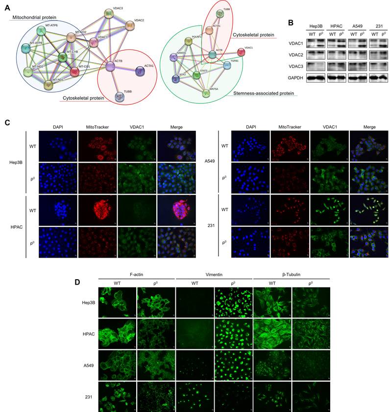
We therefore examined the expression of VDAC1, VDAC2, and VDAC3 in multiple ρ0 cell lines by western blotting. Among the three VDAC isoforms, only VDAC1 was consistently upregulated in all ρ0 cell lines, whereas the expression patterns of VDAC2 and VDAC3 varied depending on the cell line (Figure 3B). These results identified VDAC1 upregulation as a common molecular feature associated with mitochondrial dysfunction and prompted us to focus on VDAC1 in subsequent analyses.
To determine the subcellular localization of VDAC1 in ρ0 cells, immunofluorescence staining was performed using MitoTracker and an anti-VDAC1 antibody. VDAC1 staining overlapped with mitochondrial staining, indicating mitochondrial localization in both WT and ρ0 cells (Figure 3C).
To determine whether the common morphological changes induced by mitochondrial dysfunction were associated with cytoskeletal remodeling, we examined representative components of the three major cytoskeletal systems: actin for microfilaments, vimentin for intermediate filaments, and β-tubulin for microtubules. Immunofluorescence analysis showed that all ρ0 cell lines exhibited reduced actin staining intensity and altered actin organization compared with WT cells (Figure 3D). Vimentin staining intensity was increased in most ρ0 cell lines, with the exception of MDA-MB-231/ρ0 cells, whereas β-tubulin staining intensity was consistently decreased in all ρ0 cell lines. These findings indicate that mitochondrial dysfunction is accompanied by cytoskeletal remodeling, characterized by reduced actin and β-tubulin staining and cell line-dependent changes in vimentin staining intensity.
Collectively, these results identify VDAC1 upregulation and cytoskeletal remodeling as common features of chronically established ρ0 cells.
VBIT-4 attenuates multiple stemness-associated and aggressive phenotypes in ρ0 cells
To determine whether selected phenotypes observed in ρ0 cells are sensitive to pharmacologic inhibition with VBIT-4, we treated ρ0 cells with VBIT-4 and evaluated its effects on phenotypic and cytoskeletal features. VBIT-4 treatment markedly suppressed sphere formation in all ρ0 cell lines, as shown by sphere images and quantification of sphere-forming wells (Figure 4A). These data indicate that the sphere-forming ability of ρ0 cells is attenuated by VBIT-4 treatment.
Effects of VBIT-4 on selected stemness-associated and aggressive phenotypes in ρ0 cells. (A) VBIT-4 treatment markedly reduced sphere formation in ρ0 cells, as shown by sphere images and quantification of sphere-forming wells. (B) Effects of combined VBIT-4 and DX treatment on chemoresistance varied across ρ0 cell lines, as assessed by crystal violet assay. (C) VBIT-4 significantly attenuated wound closure in ρ0 cells in the wound-healing assay. (D) VBIT-4 treatment reduced the number of invading ρ0 cells in the Transwell assay. (E) Angiogenic activity induced by ρ0-derived conditioned media was reduced following VBIT-4 treatment, as shown by representative tube formation images and quantitative ImageJ-based analysis. (F) Immunofluorescence staining showed increased β-tubulin staining intensity, a modest reduction in actin staining intensity, and no marked change in vimentin staining intensity following VBIT-4 treatment in ρ0 cells. VB and V represent VBIT-4; - and + indicate the absence or presence of VBIT-4 treatment, respectively. 231 denotes MDA-MB-231. Data are presented as mean ± SD. ****P < 0.0001; ***P < 0.001; **P < 0.01; *P < 0.05; ns, not significant.
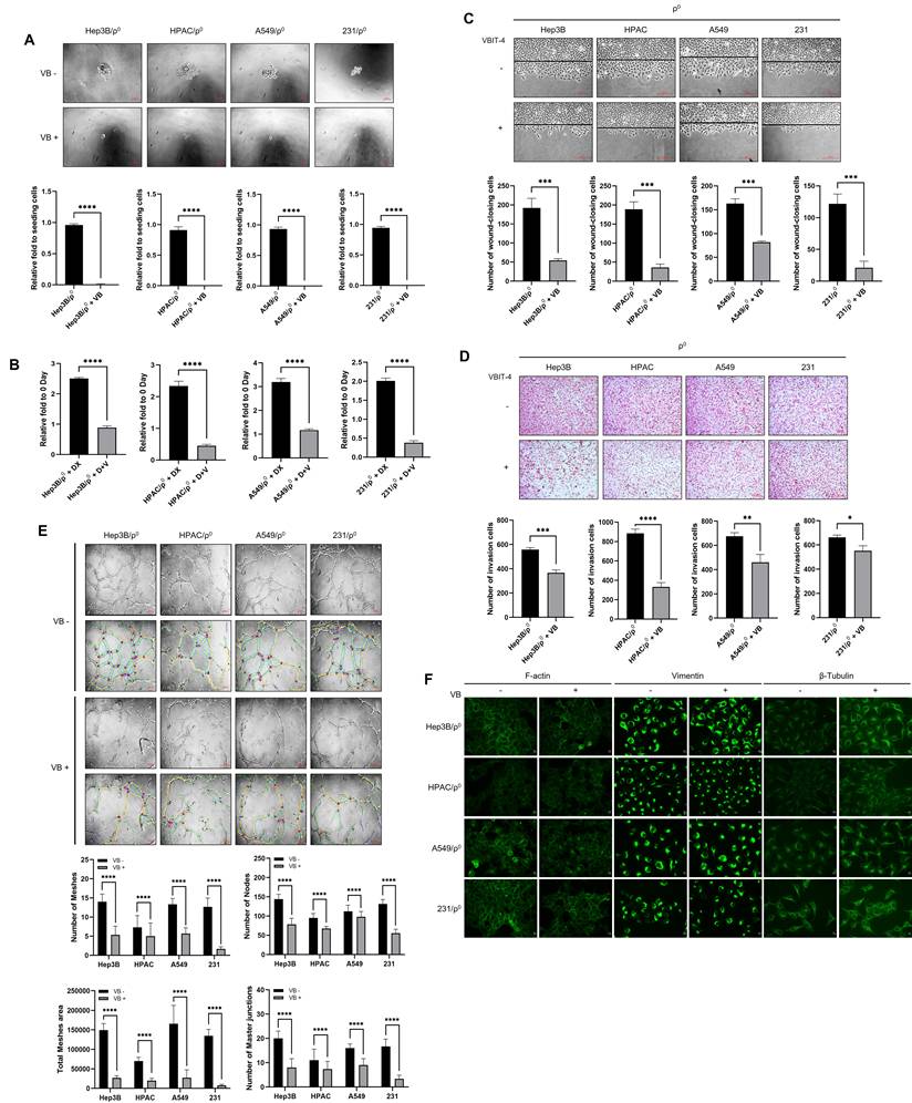
We next assessed whether VBIT-4 affects chemoresistance in ρ0 cells. The effects of combined VBIT-4 and doxorubicin treatment varied depending on the cell line. HPAC/ρ0 and MDA-MB-231/ρ0 cells exhibited a more pronounced reduction in chemoresistance following combined treatment, whereas Hep3B/ρ0 and A549/ρ0 cells exhibited comparatively more limited responses under the same conditions (Figure 4B). These findings suggest that the effects of VBIT-4 on chemoresistance-related responses are cell line-dependent and are not uniformly observed across all ρ0 models.
Because wound closure and invasion were enhanced in ρ0 cells, we further examined whether these phenotypes were affected by VBIT-4 treatment. Wound-healing assays showed that VBIT-4 treatment significantly reduced wound closure in ρ0 cells (Figure 4C). Similarly, the Transwell invasion assay showed that VBIT-4 treatment reduced the number of invading ρ0 cells (Figure 4D). These results indicate that VBIT-4 attenuates wound closure and invasion-related phenotypes in ρ0 cells.
We also examined whether VBIT-4 affects the angiogenic activity induced by ρ0 cells. Tube formation assays showed that VBIT-4 treatment reduced angiogenic activity, as demonstrated by representative images and quantitative analysis using ImageJ (Figure 4E). The numbers of nodes, master junctions, and meshes, as well as the total mesh area, were all significantly reduced following VBIT-4 treatment, indicating that VBIT-4 attenuates the angiogenic activity induced by ρ0 cells.
Finally, because VDAC1 was identified together with cytoskeleton-associated proteins and ρ0 cells exhibited common cytoskeletal alterations, we examined whether VBIT-4 treatment affects cytoskeletal organization. Immunofluorescence analysis showed no marked change in vimentin staining, a modest decrease in actin staining, and an increase in β-tubulin staining in ρ0 cells following VBIT-4 treatment (Figure 4F). These findings suggest that VBIT-4 treatment is accompanied by selective changes in cytoskeletal features, particularly in β-tubulin. Collectively, these results indicate that VBIT-4 treatment attenuates several stemness-associated and aggressive phenotypes of ρ0 cells, although its effects on chemoresistance-related responses appear to be cell line-dependent.
Discussion
Although mitochondrial dysfunction is recognized as a hallmark of cancer, its contribution to cancer cell plasticity remains incompletely understood [4, 5]. In the present study, we established mtDNA-depleted (ρ0) models from pancreatic, lung, and breast cancer cell lines and found that these cells consistently exhibited common structural features together with multiple stemness-associated and aggressive phenotypes. These findings extend our previous observations in Hep3B cells and suggest that chronically established ρ0 cells exhibit shared stemness-associated and aggressive phenotypes across multiple cancer types [8, 18].
The first notable finding of this study was that ρ0 cells exhibited common structural and growth-related changes despite their distinct tissue origins. All ρ0 cell lines exhibited complete loss of mitochondrial gene expression and reduced mitochondrial content, confirming successful establishment of the ρ0 models. These observations suggest that chronically established ρ0 cells converge toward a common cellular architecture despite differences in tissue origin. Together with the increase in proliferation, this supports the notion that chronically established ρ0 cells converge on a common cellular state that is not restricted by the tissue background of the parental cancer cells.
Chronically established ρ0 cells also exhibited multiple stemness-associated and aggressive phenotypes. In particular, all ρ0 cell lines formed spheres from single cells, indicating enhanced sphere-forming ability that was unlikely to result from bulk aggregation. The Wnt-related changes were not completely uniform across the cell lines: HPAC/ρ0 cells exhibited increased Wnt3 expression, whereas the other ρ0 models generally exhibited decreased Wnt3 and increased Wnt5 expression. In addition, ρ0 cells exhibited reduced apoptotic responses to doxorubicin, increased ABCB1 expression, enhanced wound closure and invasion, and increased angiogenic activity. Together, these results support a broader shift in chronically established ρ0 cells toward a more aggressive and phenotypically plastic state [8, 18].
Among the molecular changes identified in ρ0 cells, VDAC1 was consistently upregulated across all tested ρ0 cell lines. STRING-based analysis identified associations between VDAC family proteins and mitochondrial, cytoskeletal, and stemness-associated proteins. Among the three VDAC isoforms, only VDAC1 exhibited consistent upregulation in all ρ0 cell lines. Because VDAC1 is involved in mitochondrial permeability, metabolite transport, and apoptotic signaling, its upregulation may be associated with the chronically established ρ0 state [13, 14]. Notably, VDAC1 expression was increased despite reduced mitochondrial content, indicating that this change is not simply attributable to increased mitochondrial abundance.
In parallel with VDAC1 upregulation, all ρ0 cell lines exhibited common cytoskeletal remodeling. Actin and β-tubulin staining intensities were reduced in all ρ0 cell lines, whereas changes in vimentin staining were cell line-dependent. In particular, MDA-MB-231/ρ0 cells exhibited reduced vimentin staining, in contrast to the increased vimentin staining observed in the other ρ0 models, indicating that this component of the remodeling response was not uniform across all cell lines. These alterations were accompanied by convergence toward a similar cellular morphology, suggesting an association between mitochondrial dysfunction and shared structural remodeling in cancer cells. Although the present study does not establish a direct mechanistic link between mitochondrial dysfunction and cytoskeletal regulation, the coordinated changes in VDAC1, cellular morphology, and cytoskeletal organization suggest that these events are closely associated [16, 17].
Treatment with VBIT-4 attenuated multiple phenotypes observed in ρ0 cells. Sphere formation, wound closure, invasion, and angiogenic activity were all reduced following VBIT-4 treatment. In contrast, the effect of VBIT-4 on chemoresistance was cell line-dependent, indicating that not all phenotypes associated with the ρ0 state are regulated in the same manner. Together, these findings suggest that several phenotypes associated with the ρ0 state are sensitive to VBIT-4 treatment.
VBIT-4 treatment was also accompanied by selective cytoskeletal changes, particularly increased β-tubulin staining intensity and a modest reduction in actin staining, whereas vimentin staining was not markedly altered. These observations suggest that VBIT-4 treatment is accompanied by changes in selected structural features of ρ0 cells. In particular, β-tubulin staining intensity increased following VBIT-4 treatment; however, the relationship between this change and the common morphology of ρ0 cells remains to be clarified.
This study has several limitations. Because the experiments were performed using chronically established ρ0 cells, the findings should be interpreted in the context of cellular remodeling associated with long-term mtDNA depletion. In addition, although sphere-forming ability was consistently observed, additional functional assays assessing self-renewal capacity were not performed. Finally, although VBIT-4 treatment attenuated several phenotypes in ρ0 cells, the relationships among the ρ0 state, VDAC1 upregulation, and cytoskeletal remodeling remain to be further defined. Further studies, including in vivo validation, are needed to extend these findings.
In conclusion, chronically established ρ0 cancer cells from different tissue origins showed common structural changes together with multiple stemness-associated and aggressive phenotypes. These changes were accompanied by VDAC1 upregulation and cytoskeletal remodeling, and several of these phenotypes were attenuated by VBIT-4 treatment. Our findings suggest that VDAC1 upregulation is associated with the chronically established ρ0 state and that several phenotypes observed in ρ0 cells are sensitive to VBIT-4 treatment.
Abbreviations
mtDNA: mitochondrial DNA; VDACs: voltage-dependent anion channels; DMEM: Dulbecco's modified Eagle's medium; FBS: fetal bovine serum; DX: doxorubicin; HUVEC: human umbilical vein endothelial cell; RT-PCR: reverse transcriptase-polymerase chain reaction; CM: conditioned medium; WT: wild-type; SD: standard deviation; ANOVA: analysis of variance.
Acknowledgements
This work was supported by a National Research Foundation of Korea (NRF) grant funded by the Korean government (MOE) (RS-2023-00242974) and a grant from the Center for Women in Science, Engineering and Technology (WISET) (2025-275).
Data availability
The data generated and/or analyzed during the current study are available from the corresponding author on reasonable request.
Competing Interests
The authors have declared that no competing interest exists.
References
1. Giacomello M, Pyakurel A, Glytsou C, Scorrano L. The cell biology of mitochondrial membrane dynamics. Nat Rev Mol Cell Biol. 2020;21:204-24
2. Liu BH, Xu CZ, Liu Y, Lu ZL, Fu TL, Li GR. et al. Mitochondrial quality control in human health and disease. Mil Med Res. 2024;11:32
3. Suomalainen A, Battersby BJ. Mitochondrial diseases: the contribution of organelle stress responses to pathology. Nat Rev Mol Cell Biol. 2018;19:77-92
4. Wang SF, Tseng LM, Lee HC. Role of mitochondrial alterations in human cancer progression and cancer immunity. J Biomed Sci. 2023;30:61
5. Liu Y, Sun Y, Guo Y, Shi X, Chen X, Feng W. et al. An overview: the diversified role of mitochondria in cancer metabolism. Int J Biol Sci. 2023;19:897-915
6. Awad AMAM, Abdul Karim N. Dysregulation of mitochondrial function in cancer cells. Int J Mol Sci. 2025;26:6750
7. Du H, Xu T, Yu S, Wu S, Zhang J. Mitochondrial metabolism and cancer therapeutic innovation. Signal Transduct Target Ther. 2025;10:245
8. Han YS, Yi EY, Jegal ME, Kim YJ. Cancer stem-like phenotype of mitochondria dysfunctional Hep3B hepatocellular carcinoma cell line. Cells. 2021;10:1608
9. Tan EW, Singh SK, Dua K, Gupta G, Lee WL, Wong RSY. et al. Cancer stem cells: mitochondria signalling pathway and strategies for therapeutic interventions. Mol Biol Rep. 2025;52:671
10. Nie X, Liu H, Ye W, Wei X, Fan L, Ma H. et al. LRP5 promotes cancer stem cell traits and chemoresistance in colorectal cancer. J Cell Mol Med. 2022;26:1095-112
11. Wu M, Zhang X, Zhang W, Chiou YS, Qian W, Liu X. et al. Cancer stem cell regulated phenotypic plasticity protects metastasized cancer cells from ferroptosis. Nat Commun. 2022;13:1371
12. Yadav AK, Desai NS. Cancer stem cells: acquisition, characteristics, therapeutic implications, targeting strategies and future prospects. Stem Cell Rev Rep. 2019;15:331-55
13. Huang H, Chen M, Feng S, Lin Z, Liu Y. The dual role of VDAC in cancer: molecular mechanisms and advances in targeted therapy. Biomed Pharmacother. 2025;191:118530
14. Camara AKS, Zhou Y, Wen PC, Tajkhorshid E, Kwok WM. Mitochondrial VDAC1: a key gatekeeper as potential therapeutic target. Front Physiol. 2017;8:460
15. Argueti-Ostrovsky S, Barel S, Kahn J, Israelson A. VDAC1: a key player in the mitochondrial landscape of neurodegeneration. Biomolecules. 2025;15:33
16. Puurand M, Tepp K, Timohhina N, Aid J, Shevchuk I, Chekulayev V. et al. Tubulin betaII and betaIII isoforms as the regulators of VDAC channel permeability in health and disease. Cells. 2019;8:239
17. Ji Y, Lin Y, He J, Xie Y, An W, Luo X. et al. Research progress of mitochondria and cytoskeleton crosstalk in tumour development. Biochim Biophys Acta Rev Cancer. 2025;1880:189254
18. Yi EY, Park SY, Jung SY, Jang WJ, Kim YJ. Mitochondrial dysfunction induces EMT through the TGF-beta/Smad/Snail signaling pathway in Hep3B hepatocellular carcinoma cells. Int J Oncol. 2015;47:1845-53
Author contact
Corresponding author: Yung-Jin Kim, Department of Molecular Biology, Pusan National University, Busan 46241, Republic of Korea. Phone Number: +82-10-4149-2935; E-mail: yjinkimac.kr.