Impact Factor ISSN: 1837-9664




J Cancer 2026; 17(9):1600-1614. doi:10.7150/jca.130206 This issue Cite
Review
Overcoming Resistance to PARP Inhibitors in BRCA-Mutated Cancers: Mechanisms and Therapeutic Strategies
Jiahui Guo1,2*, Yilei Wang3*, Jie Li1  , Zhitao Jiang1
1. Department of Pharmacy, Zhangjiagang TCM Hospital, Affiliated to Nanjing University of Chinese Medicine, Zhangjiagang, Jiangsu, 215600, China.
2. School of Medicine, Nanjing University of Chinese Medicine, Nanjing, Jiangsu, 210023, China.
3. Department of Pharmacy, Taicang TCM Hospital, Affiliated to Nanjing University of Chinese Medicine, Taicang, Jiangsu, 215400, China.
*These authors contributed equally to this work.
Received 2025-12-19; Accepted 2026-8-24; Published 2026-9-3
Abstract
The tumor suppressor genes BRCA1 and BRCA2 are essential for homologous recombination (HR) repair. Mutations in BRCA1/2 compromise HR repair efficacy, resulting in heightened susceptibility of tumor cells to poly (ADP-ribose) polymerase inhibitors (PARPi) via synthetic lethality. To date, several PARPi have been approved for BRCA-mutated and HR-deficient tumors, but acquired resistance and cumulative toxicity continue to limit their long-term clinical benefit. This narrative review offers an updated mechanistic model of PARPi action and categorizes resistance mechanisms by clinical relevance and evidentiary strength. Importantly, we distinguish between standard combination therapies in the PARPi-naïve setting and interventions explicitly tailored for PARPi-exposed or PARPi-resistant disease. Building on this distinction, we propose a biomarker-guided framework that links specific resistance phenotypes to appropriate specimen types, detection assays, and candidate interventions. Through this evidence-based clinical framework, we aim to clarify the current therapeutic landscape, improve data interpretation, and guide prospective trial design.
Keywords: PARP inhibitors, BRCA1/2 mutations, synthetic lethality, tumor therapy, PARPi resistance
1. Introduction
DNA damage is generated by endogenous metabolic processes and by exogenous agents, including chemicals, ionizing radiation, and ultraviolet light [1-5]. Lesions range from base mismatches and DNA crosslinks to single-strand breaks (SSBs) and double-strand breaks (DSBs) [6]. Although an SSB does not immediately disrupt both strands of the DNA duplex, an unrepaired SSB encountered by a replication fork may generate a single-ended or double-ended DSB [7, 8]. DSBs are particularly cytotoxic because inaccurate repair can produce deletions, translocations, and other chromosomal abnormalities that compromise genomic stability [5, 9, 10]. Major DSB repair pathways include nonhomologous end joining (NHEJ), homologous recombination (HR), single-strand annealing (SSA), and microhomology-mediated end joining (MMEJ) [11-13]. NHEJ directly ligates DNA ends throughout the cell cycle and is often error-prone, whereas polymerase theta (Polθ)-dependent MMEJ uses short microhomologies, typically 2-20 nucleotides [13-21]. HR is largely restricted to the S and G2 phases, uses a homologous template, usually the sister chromatid, and is therefore a high-fidelity repair pathway [22]. BRCA1 promotes repair pathway choice and DNA end resection, whereas 53BP1-dependent end protection favors NHEJ; the balance between these activities is a key determinant of repair outcome [23-25].
BRCA1 and BRCA2 were identified in the 1990s as major tumor suppressor genes, and pathogenic variants in both genes increase the risk of breast, ovarian, and several other cancers [26, 27]. Although the two proteins cooperate in HR, their functions are not identical. BRCA1 acts mainly upstream by coordinating checkpoint signaling, promoting DNA-end resection, and directing lesions toward HR during S/G2 [28, 29]. BRCA2 acts downstream by loading and stabilizing RAD51 on single-stranded DNA (ssDNA), enabling homology search and strand invasion [30, 31]. Both proteins also participate in the response to replication stress, but BRCA2 has a particularly direct role in stabilizing RAD51 filaments and protecting nascent DNA at stalled replication forks. These functional differences are relevant to resistance because mechanisms that partly bypass BRCA1 loss may not compensate for the loss of BRCA2-mediated RAD51 loading [32].
PARP1 detects DNA lesions, promotes local chromatin remodeling, and organizes repair through poly(ADP-ribosyl)ation. PARPi inhibit catalytic activity and can stabilize PARP1-DNA complexes, thereby increasing replication-associated damage in HR-deficient cells [33-37]. The term BRCAness is more appropriately used for non-BRCA tumors that phenocopy selected features of BRCA deficiency and should not be treated as synonymous with a germline BRCA mutation [38, 39]. The resulting synthetic lethal interaction preferentially affects tumor cells that have lost both functional BRCA alleles, whereas most normal tissues in germline mutation carriers retain one wild-type allele and greater HR capacity [40-44]. PARPi have produced substantial clinical benefit in several BRCA1/2-mutated cancers, but resistance, toxicity, and differences among tumor types and treatment settings limit their effectiveness [45-47]. This review therefore summarizes BRCA1/2 biology, updates the mechanistic basis of PARP inhibition, ranks resistance mechanisms by clinical relevance and evidentiary strength, and evaluates biomarker-guided strategies for PARPi-exposed or PARPi-resistant disease.
1.1 Scope and evidence-synthesis approach
This article is a narrative review focused primarily on cancers carrying pathogenic germline or somatic BRCA1/2 alterations. Evidence from non-BRCA HR deficiency, BRCAness, other DNA repair defects, or genotype-independent combinations is included only when it clarifies PARPi mechanism, resistance, biomarker performance, or therapeutic generalizability; these categories are not treated as biologically interchangeable. A focused search of PubMed/MEDLINE and ClinicalTrials.gov was conducted for English-language reports published from January 2005 onward using terms related to PARP inhibition, BRCA1/2, HR, resistance, reversion, replication forks and gaps, transcription-replication conflicts, ctDNA, RAD51 foci, biomarkers, and relevant agents or trials. Official FDA, EMA, and NMPA sources were consulted in the revised evidence synthesis for regulatory-status information. Evidence was interpreted qualitatively as regulatory or randomized clinical evidence, prospective or longitudinal patient evidence, patient-derived translational evidence, or mechanistic cell-line/animal evidence. Because the aim was conceptual and translational synthesis, formal meta-analysis and risk-of-bias scoring were not performed.
2. BRCA1/2 and HR Repair
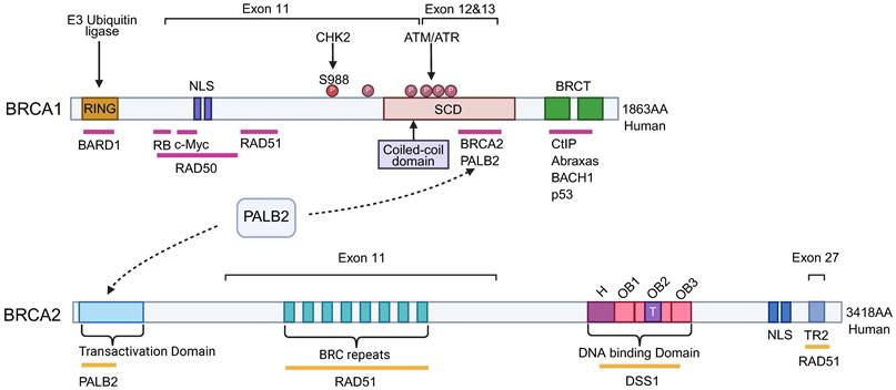
BRCA1 is located on chromosome 17 and contains 22 coding exons spanning approximately 100 kb of genomic DNA [40]. It encodes a 1,863-amino-acid protein of approximately 220 kDa that participates in DNA damage repair, cell-cycle control, and transcription [48-50]. BRCA1 contains two nuclear localization signals and three major functional regions: an N-terminal RING domain, a central coiled-coil (CC) domain, and tandem C-terminal BRCT repeats. The RING domain forms an obligate heterodimer with BARD1 and contributes to E3 ubiquitin-ligase activity and damage-site recruitment [51-53]. A serine cluster domain within exons 11-13 contains multiple ATM/ATR-responsive phosphorylation sites that help coordinate checkpoint signaling and localization after DNA damage [54]. The CC domain binds PALB2 and links BRCA1 to the PALB2-BRCA2-RAD51 axis [55]. The tandem BRCT domains, separated by a short linker, recognize phosphorylated partners such as CtIP, Abraxas, and FANCJ/BACH1/BRIP1; pathogenic variants in this region can disrupt substrate recognition, localization, and checkpoint function [56-59] (Figure 1 and Table 1).
Functional Domains of BRCA1 and BRCA2
| Gene | Domain | Residue range | Principal functional roles and mechanisms |
|---|---|---|---|
| BRCA1 | RING domain | 24-65 | Coordinates zinc ions and forms an obligate heterodimer with BARD1, supporting E3 ubiquitin-ligase activity, DNA-damage signaling, and recruitment to damaged chromatin [51, 52, 176]. |
| NLS | 501-507 and 607-614 | Provide binding interfaces for importin-alpha and support active nuclear import of BRCA1-containing complexes [177, 178]. | |
| Serine Cluster Domain (SCD) | 1280-1524 | Contains multiple ATM/ATR-responsive phosphorylation sites, including Ser1387 and Ser1524, that coordinate localization and S-phase/G2-M checkpoint signaling after DNA damage [54]. | |
| Coiled-coil (CC) domain | 1364-1437 | Binds PALB2 and promotes assembly of the BRCA1-PALB2-BRCA2 repair complex [55, 82]. | |
| BRCT Repeat Domains | 1650-1863 | Tandem phosphoprotein-binding repeats recognize phosphorylated CtIP, Abraxas, and FANCJ and coordinate DNA-end resection and damage signaling [56, 57, 59, 179]. | |
| BRCA2 | N-terminal Binding Region | 21-44 | Binds PALB2 and EMSY, linking upstream DNA damage signaling to HR and chromatin-regulatory networks [55, 82, 180]. |
| BRC Repeats (1 through 8) | ~1000-2085 | Eight conserved repeats within exon 11 interact with RAD51, promote RAD51 recruitment to ssDNA, and regulate RAD51 binding to double-stranded DNA [67, 70]. | |
| DNA-Binding Domain (DBD) | 2478-3185 | Oligonucleotide-binding folds engage ssDNA and the tower domain contacts double-stranded DNA; DSS1 stabilizes the domain and supports HR and interstrand-crosslink repair [68, 69]. | |
| C-terminal RAD51-Binding Domain (TR2) | Exon 27 (includes S3291) | Provides a second RAD51 interaction site, stabilizes mature RAD51 filaments, and protects stalled replication forks. CDK-dependent S3291 phosphorylation regulates this activity during the cell cycle [70, 71, 181]. | |
| NLS | C-terminal basic clusters | Direct nuclear import of the large BRCA2 complex; loss of these signals impairs nuclear localization and repair function [182]. |
Main Functional Domains of BRCA1 and BRCA2. BRCA1 comprises 1,863 amino acids and contains an N-terminal RING domain, two nuclear localization signals (NLSs), a serine cluster domain (SCD), a coiled-coil (CC) domain, and tandem C-terminal BRCT repeats. The RING domain interacts with BARD1; the SCD spans exons 11-13 and contains multiple ATM/ATR-responsive phosphorylation sites; and the BRCT repeats recognize phosphorylated partners including CtIP, Abraxas, and BACH1. BRCA2 comprises 3,418 amino acids. Exon 11 contains eight conserved BRC repeats that mediate RAD51 interactions. The N-terminal region binds PALB2 and maintains the BRCA1-PALB2-BRCA2 axis. The C-terminal region contains nuclear localization signals and an additional RAD51-binding site encoded by exon 27. The DNA-binding domain (DBD) comprises an alpha-helical region, three oligonucleotide-binding (OB) folds, and a tower domain; it interacts with DSS1 to support HR repair.
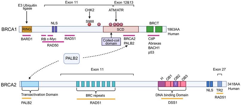
BRCA2 is located on chromosome 13, contains 27 coding exons, and encodes a 3,418-amino-acid protein [60]. Exon 11 contains eight conserved BRC repeats that interact with RAD51, promote RAD51 loading onto ssDNA, and regulate its association with double-stranded DNA [61-63] (Figure 1). The N-terminal region binds PALB2 and maintains the BRCA1-PALB2-BRCA2 repair axis [54, 64]. The C-terminal region contains nuclear localization signals and an additional RAD51-binding site encoded by exon 27; phosphorylation of S3291 by cyclin-dependent kinases regulates this interaction and contributes to replication-fork protection [65, 66]. The DNA-binding domain includes an alpha-helical region, three oligonucleotide-binding folds, and a tower domain, allowing BRCA2 to engage both ssDNA and double-stranded DNA. DSS1 stabilizes BRCA2, promotes its nuclear localization, and facilitates RAD51 loading onto RPA-coated ssDNA [67-69]. Beyond canonical HR, BRCA2 suppresses post-replicative ssDNA gaps, protects nascent DNA from nuclease-mediated degradation, and contributes to cytokinesis and chromosomal stability [70-73].
HR repairs DSBs and replication-associated lesions by using an intact homologous template [74]. BRCA1 and BRCA2 are central regulators of homology-directed repair, and functional disruption of either protein can compromise genome maintenance and therapy response [75, 76]. BRCA1 functions upstream in this pathway through interactions with phosphorylated CtIP and the MRE11-RAD50-NBS1 (MRN) complex. These interactions promote 5′ to 3′ DNA end resection and generate the 3′ ssDNA overhang required for RAD51 filament formation [58, 77, 78]. CDK-dependent phosphorylation of CtIP facilitates BRCA1 binding and limits RIF1-dependent end protection. In parallel, the BRCA1-BARD1 heterodimer modifies chromatin near DSBs and antagonizes 53BP1, thereby restricting rapid but potentially inaccurate NHEJ and favoring HR [79]. This upstream role explains why loss of 53BP1-RIF1-Shieldin can partially restore HR in some BRCA1-deficient models.
BRCA2 acts downstream to assemble and stabilize the RAD51 nucleoprotein filament. After resection, replication protein A (RPA) protects ssDNA from nucleases. PALB2 bridges BRCA1 and BRCA2 at damaged chromatin, after which BRCA2 replaces RPA with RAD51 and stabilizes the resulting RAD51-ssDNA filament [65, 78]. RAD51 then searches for a homologous sequence on the sister chromatid and promotes strand invasion and D-loop formation. DSS1 assists this process by weakening RPA-ssDNA interactions and stabilizing the BRCA2-DSS1-RAD51 complex [80]. The PALB2-BRCA2-RAD51 axis favors high-fidelity HR and limits mutagenic alternatives such as RAD52-dependent SSA [81, 82]. BRCA2 also helps restrain inappropriate end joining and protects stalled replication forks. Therefore, restoration of DNA end resection alone generally cannot compensate for BRCA2 deficiency [83].
3. Antitumor Mechanisms of PARPi
ADP-ribosylation is a reversible post-translational modification that uses nicotinamide adenine dinucleotide (NAD+) as the donor of ADP-ribose units [84, 85]. Attachment of one unit is termed mono(ADP-ribosyl)ation, whereas polymer formation is termed poly(ADP-ribosyl)ation (PARylation). In humans, these reactions are catalyzed by the PARP/ADP-ribosyltransferase family, whose members regulate cellular stress responses, chromatin structure, DNA repair, and cell death [10, 86, 87]. PARP1, PARP2, and PARP3 are DNA-damage-responsive family members, with PARP1 accounting for most damage-induced PAR synthesis and therefore representing the best-characterized therapeutic target [88]. PARP2 shares substantial similarity with the PARP1 catalytic domain but lacks the N-terminal zinc-finger architecture. Their partially overlapping functions and the embryonic lethality associated with combined loss emphasize the importance of PAR metabolism in genome maintenance [89].
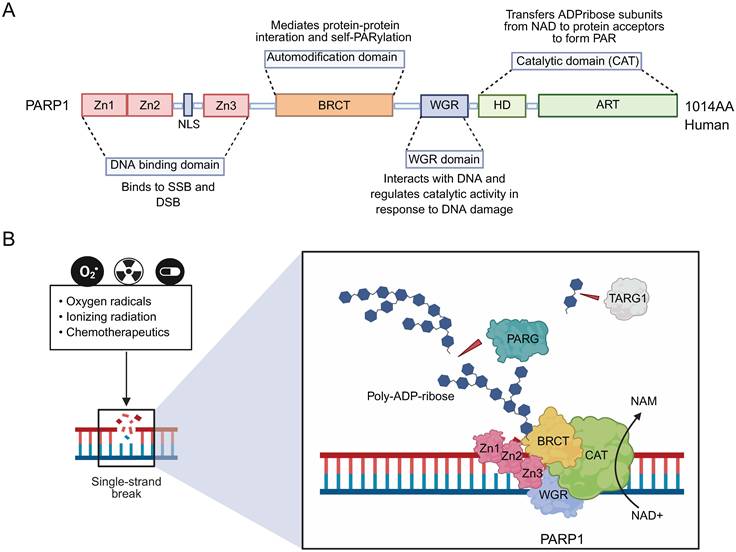
PARP1 was initially characterized as a factor in SSB repair but is now recognized as a multifunctional regulator of several DNA repair and chromatin-remodeling processes [90]. Its N-terminal Zn1 and Zn2 domains recognize DNA discontinuities, whereas Zn3 and the WGR domain transmit the DNA-binding signal to the catalytic region. A nuclear localization signal lies between Zn2 and Zn3, and the central automodification region provides major sites for protein interaction and auto-PARylation. The C-terminal catalytic domain contains a helical domain and an ADP-ribosyltransferase domain that binds NAD+ and synthesizes PAR [91-94] (Figure 2A). DNA binding induces a conformational change that activates catalysis, producing a dense, negatively charged PAR scaffold that promotes chromatin relaxation and the recruitment or organization of repair factors [95-100]. Extensive auto-PARylation facilitates PARP1 release from DNA. PARG then hydrolyzes PAR chains, and TARG1 removes terminal mono(ADP-ribose), maintaining a dynamic cycle of PAR synthesis and degradation [101, 102] (Figure 2B).
PARP1 Domains and PAR Metabolism Relevant to PARP Inhibition. (A) PARP1 contains an N-terminal DNA-binding region with Zn1, Zn2, and Zn3 zinc-finger domains, an NLS between Zn2 and Zn3, a central automodification region containing the BRCT domain, a WGR domain, and a C-terminal catalytic (CAT) domain composed of the helical (HD) and ADP-ribosyltransferase (ART) domains. (B) After binding a DNA nick or break, PARP1 uses NAD+ to synthesize PAR. Extensive auto-PARylation promotes PARP1 release from DNA through electrostatic repulsion. PARG degrades PAR chains, whereas TARG1 removes terminal mono(ADP-ribose), maintaining dynamic PAR turnover.
PARPi exploit the dependence of BRCA1/2-deficient cells on PARP1-mediated responses to replication-associated damage [103]. Catalytic inhibition reduces PAR synthesis and impairs repair-factor organization, whereas inhibition of auto-PARylation can prolong the residence of PARP1 at DNA lesions, a process commonly termed PARP1 trapping [18, 104, 105]. The traditional model proposes that unrepaired SSBs are converted into DSBs during replication and become lethal when HR is defective. This explanation remains useful but is not sufficient as a universal linear pathway. PARPi can also promote post-replicative ssDNA gaps, abnormal fork remodeling, fork stress, and transcription-replication conflicts. PARP1, TIMELESS, and TIPIN help protect replisomes during such conflicts. In BRCA-deficient cells, unresolved conflicts can generate persistent DNA damage and abnormal mitotic chromatin [106]. Thus, catalytic inhibition, trapping, ssDNA-gap accumulation, replication stress, and transcription-replication conflicts should be viewed as parallel and interacting sources of cytotoxicity whose relative contributions vary with the drug, BRCA genotype, and cellular context.
PARPi have demonstrated clinical activity in selected ovarian, breast, pancreatic, and prostate cancers, but regulatory indications differ by jurisdiction, biomarker requirement, and treatment setting and continue to evolve [107-114]. Olaparib, rucaparib, niraparib, talazoparib, fuzuloparib, and pamiparib illustrate the diversity of approved agents and regional labels; fixed statements about a single worldwide number of approved PARPi should therefore be avoided. Olaparib has generated pivotal evidence in BRCA-mutated ovarian, breast, pancreatic, and prostate cancers [108-110], whereas other agents have setting-specific roles, including ovarian maintenance, BRCA-mutated breast cancer, and selected regional indications [111-114]. Importantly, benefit in a first-line or maintenance trial does not demonstrate reversal of established PARPi resistance. Table 2 is retained as a historical overview rather than a substitute for current FDA, EMA, NMPA, or other local prescribing information. Accordingly, approved indications and PARPi-naïve treatment strategies should be distinguished from the investigational resistance-specific approaches discussed in Section 5.
Selected Historical Milestones in PARP Biology and PARP Inhibitor Development. Regulatory milestones are shown for historical context and do not imply efficacy in established PARPi-resistant disease.
| Year | Events | Summary |
|---|---|---|
| 1960s | Discovery of PAR and PARylation | Pierre Chambon and colleagues identified NAD+-dependent PARylation, establishing the foundation for PARP research [183]. |
| 1970s-1980s | Discovery of PARG and expansion of PARP1 biology | PARG was identified, and the roles of PARP1 in PAR signaling, chromatin remodeling, metabolism, cell death, and DNA repair were progressively defined [184-186]. |
| 1990s | Identification of BRCA1/2 and characterization of PARP family structure | BRCA1/2 were established as tumor-suppressor genes required for HR, while the domains and functions of PARP family members were increasingly characterized [187, 188]. |
| 2005 | Demonstration of synthetic lethality between PARP inhibition and BRCA deficiency | Independent studies showed that BRCA1/2-deficient cells are selectively sensitive to PARP inhibition, establishing a therapeutic synthetic-lethal strategy [43]. |
| 2009 | Phase I clinical development of olaparib | Early clinical testing demonstrated antitumor activity with a manageable safety profile, providing proof of concept for PARPi in patients [189]. |
| 2014 | First regulatory approval of olaparib | The FDA approved olaparib for a BRCA-associated ovarian cancer setting, marking the clinical translation of PARP-BRCA synthetic lethality [108, 109]. |
| 2016 | Regulatory approval of rucaparib | Rucaparib became another FDA-approved PARPi in ovarian cancer [111]. |
| 2017 | Regulatory approval of niraparib | Niraparib expanded PARPi maintenance treatment in ovarian cancer [112]. |
| 2018 | Regulatory approval of talazoparib | Talazoparib was approved for HER2-negative advanced or metastatic breast cancer with germline BRCA mutations [113]. |
| 2021 | Regional approvals of fuzuloparib and pamiparib | The NMPA approved fuzuloparib and pamiparib in selected BRCA-mutated ovarian-cancer settings [114]. |
| 2022 | Clinical development of selective PARP1 inhibition | Early clinical results for the selective PARP1 inhibitor AZD5305 supported further evaluation of a potentially wider therapeutic index [116]. |
| 2024 | Greater recognition of transcription-replication conflicts | Mechanistic studies identified unresolved transcription-replication conflicts as an important source of PARPi-induced damage in HR-deficient cells, complementing rather than universally replacing trapping-based models [190]. |
The synthetic lethal activity of this drug class is driven principally by PARP1, whereas inhibition of PARP2 and other PARP family members may contribute to hematologic and other toxicities. This has encouraged the development of more selective PARP1 inhibitors. Saruparib (AZD5305) is a highly selective PARP1 inhibitor and trapper that has shown favorable target selectivity, tolerability, and preliminary activity in early clinical development [115, 116]. However, saruparib remains investigational. It should not be described as an approved treatment or as a proven means of overcoming a specific resistance mechanism, although its wider therapeutic index may support future combination strategies and evaluation in PARPi-exposed populations.
4. Mechanisms of PARPi Resistance
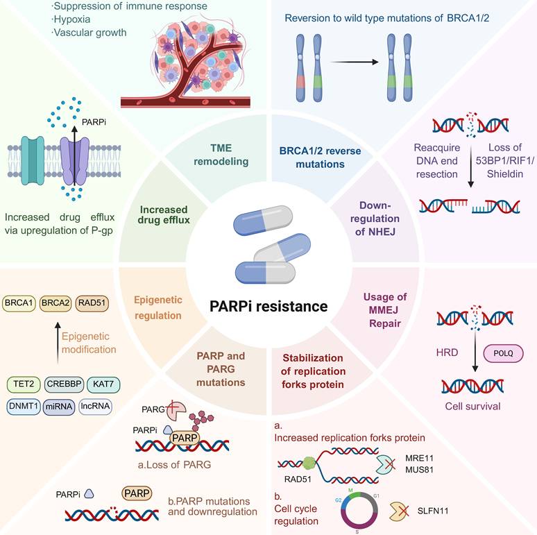
PARPi resistance may be intrinsic, adaptive, or acquired. Intrinsic resistance refers to the absence of meaningful initial benefit despite adequate drug exposure, whereas acquired resistance follows a period of clinical response or disease control [117]. A fixed duration threshold is not biologically universal and should not replace trial-specific definitions. Adaptive drug tolerance describes a reversible or semi-stable state in which cells survive treatment without an immediately identifiable genetic driver. The proposed progression from a tolerant persister state to stable genetic escape is a useful evolutionary model, but it is not obligatory: tumors may contain pre-existing resistant clones, acquire several mechanisms simultaneously, retain non-genetic resistance, or develop different mechanisms at separate metastatic sites [118, 119]. For clinical interpretation, the mechanisms discussed below can be viewed in a practical hierarchy. BRCA1/2 reversion and restoration of HR have the strongest patient-level support; fork protection, altered PARP/PARG biology, and drug efflux are translationally supported but context-dependent; and epigenetic or microenvironmental remodeling remains predominantly preclinical (Figure 3).
Mechanisms of PARPi Resistance. Major mechanisms include BRCA1/2 reversion and restoration of HR, loss of end protection, replication-fork stabilization, altered PARP/PARG biology, increased drug efflux, epigenetic regulation, and tumor microenvironment remodeling. BRCA1/2 reversion and HR restoration have the strongest patient-level support; fork protection, altered PARP/PARG biology, and drug efflux are translationally supported but context-dependent and epigenetic or microenvironmental remodeling remains predominantly preclinical.
The relative contribution of each resistance pathway varies with tumor type, disease stage, prior platinum and PARPi exposure, hormonal signaling, and the timing and method of sampling [120-124]. BRCA reversion mutations are well documented in ovarian, breast, pancreatic, and prostate cancers, but their apparent prevalence is cohort-specific. Advanced and heavily pretreated tumors often contain greater clonal diversity and may harbor several concurrent mechanisms [122]. Androgen receptor signaling can intersect with DNA-repair programs in prostate cancer, whereas estrogen receptor signaling may influence BRCA and HR-gene expression in breast and ovarian cancers [123, 124]. These observations support a tumor- and context-specific interpretation rather than a universal sequence of resistance events.
4.1 Restoration of HR Repair
Restoration of BRCA function and HR is the best-established acquired resistance mechanism in BRCA1/2-mutated cancers. Secondary substitutions, insertions, deletions, or structural events may restore the BRCA1/2 open reading frame or generate a partially functional protein, thereby reducing synthetic lethality under platinum or PARPi selection [125, 126]. Reversion mutations have been observed after treatment in multiple BRCA-associated tumor types and may arise in several convergent subclones. Longitudinal circulating tumor DNA (ctDNA) can detect some reversion clones before or at radiographic progression and can capture spatial heterogeneity that a single biopsy may miss [118]. Nevertheless, a negative plasma result does not exclude resistance because tumor shedding and assay sensitivity vary. Tissue and plasma results should therefore be interpreted together when feasible, and the detection of a reversion does not yet mandate a standardized subsequent therapy. BRCA1 hypomorphic splice isoforms, including Δ11 or Δ11q forms, and HSP90-mediated stabilization of mutant BRCA1 can also restore sufficient function without a full canonical reversion.
RAD51 is central to homology search, strand invasion, and HR restoration. Downregulation of early mitotic inhibitor 1 (EMI1), a component of the ubiquitin-ligase machinery that regulates RAD51 turnover, can increase RAD51 abundance and confer PARPi resistance in BRCA1-deficient models [127]. TOPBP1-dependent phosphorylation of RAD51 at serine 14 also influences the replication-stress response, and loss of TOPBP1 can increase PARPi sensitivity [128]. More broadly, restoration of RAD51 nuclear foci provides a functional indication that HR competence has returned, regardless of whether the underlying event is a BRCA reversion, altered splicing, increased RAD51 expression, or another pathway change. RAD51-foci assays are therefore promising translational biomarkers, although preanalytical conditions, scoring thresholds, and interlaboratory standardization remain important limitations.
In BRCA1-deficient tumors, loss of end-protection factors can partially restore HR by permitting DNA end resection. Reduced activity of 53BP1, RIF1, Artemis, REV7/Shieldin, or the CST complex can therefore decrease PARPi sensitivity [129-131]. This escape route is more biologically plausible in BRCA1 deficiency than in BRCA2 deficiency, because end resection cannot by itself replace the BRCA2-dependent loading of RAD51. MicroRNAs such as miR-622 may also shift repair away from NHEJ by reducing Ku expression [132]. Polθ-mediated MMEJ is frequently used by HR-deficient tumor cells and can support survival or generate microhomology-associated reversion events [133]. However, MMEJ dependence, HR restoration, and BRCA reversion should be distinguished mechanistically rather than presented as a single equivalent process.
4.2 Stabilization of Replication Forks
Replication-fork protection can reduce PARPi sensitivity without fully restoring canonical HR. PARP1, BRCA1, and BRCA2 normally help protect stalled or reversed forks from degradation by nucleases such as MRE11 and EXO1. Loss of PTIP or CHD4 can reduce MRE11 recruitment, while loss of fork-remodeling factors such as SMARCAL1, ZRANB3, or HLTF can limit nuclease-dependent degradation of nascent DNA [134, 135]. Alterations involving SLFN11, EZH2-MUS81 signaling, DYNLL1, or RADX can also modify replication-stress tolerance and PARPi response [136-138]. These mechanisms are strongly supported in cell lines and patient-derived models, but standardized clinical assays and prospective evidence remain limited. Fork protection should therefore be regarded as a translationally supported, context-dependent mechanism rather than as clinically equivalent to BRCA reversion.
4.3 Increased Drug Efflux
Drug efflux lowers the intracellular concentration of anticancer agents and is a recognized mechanism of multidrug resistance. Amplification, promoter rearrangement, or overexpression of ABCB1/MDR1 can increase expression of the membrane transporter P-glycoprotein, which uses ATP hydrolysis to export selected drugs and reduce intracellular exposure [139]. ABCB1-mediated resistance has been described in experimental models and some clinical samples after chemotherapy or PARPi treatment. Its relevance, however, depends on whether the specific PARPi is an effective P-glycoprotein substrate. It is therefore inappropriate to assume that all PARPi have identical susceptibility to efflux, and routine clinical testing for this mechanism has not been established.
4.4 Decreased PARP Trapping
Alterations in PARP1 structure, activation, or chromatin residence can reduce effective trapping and contribute to resistance. For example, the PARP1 R591C mutation disrupts communication between the WGR and DNA-binding regions, allowing rapid dissociation from damaged DNA and reducing inhibitor-induced trapping [140]. Phosphorylation of PARP1 at Y907 by c-MET can enhance catalytic activity and reduce inhibitor binding [141]. Conversely, inhibition or loss of the deubiquitinase USP1 increases PARP1 ubiquitination and chromatin retention, thereby enhancing PARPi-induced damage in experimental models [142]. These observations provide a mechanistic rationale for USP1- or c-MET-directed combinations, but their prevalence and predictive value in patients with PARPi-resistant disease remain uncertain.
HMGB3 also regulates PARP1 dynamics. HMGB3 is highly expressed in some stem-like and cancer-cell populations and can facilitate PARP1 dissociation from chromatin; its loss or downregulation may increase PARP1 trapping and PARPi sensitivity. Other processes, including TIMELESS-TIPIN-dependent replisome protection and APEX1/KAT6A-associated phase separation, may further influence PARP1 residence at damaged chromatin [143, 144]. Most evidence for these mechanisms is preclinical. They should be presented as emerging modifiers of PARP1 trapping rather than as established clinical causes of resistance.
4.5 Loss of PARG
PARG reverses PARylation by degrading PAR chains. Loss of PARG or reduced PARG activity can preserve residual PAR signaling despite PARP inhibition, promote PARP1 release from DNA, and maintain limited recruitment of repair factors, thereby reducing PARPi cytotoxicity [145, 146]. PARG loss has been identified in resistant experimental models and selected tumor samples, but its clinical frequency and optimal detection method are not yet defined. PARG inhibitors and PARP/PARG combinations are being evaluated preclinically; these approaches remain investigational and should not be interpreted as validated strategies for patients with a detected PARG alteration.
4.6 Tumor Microenvironment Remodeling
The tumor microenvironment may modify PARPi response through hypoxia, metabolic stress, stromal signaling, and immune regulation. Moderate hypoxia can reduce reactive oxygen species and attenuate PARPi-associated DNA damage in HR-deficient models, sometimes independently of HIF signaling [147, 148]. Hypoxia-activated agents such as tirapazamine have enhanced PARPi activity in experimental systems, but this strategy has not been established clinically. The available evidence therefore supports hypoxia as a plausible, context-dependent contributor rather than a universal resistance mechanism.
PARPi can acutely increase cytosolic DNA and activate cGAS-STING and type I interferon signaling, creating a pro-immunogenic state [149]. With prolonged exposure, however, chronic inflammatory signaling may be accompanied by compensatory immunosuppression, including STAT3/IL-34 activation, regulatory T-cell expansion, and cancer-associated fibroblast-derived signals such as CCL2 [150]. These changes may protect residual tumor cells from immune clearance. Because most data derive from preclinical models or small translational studies, immune remodeling should be classified as an emerging mechanism and not as a dominant explanation for resistance in all BRCA-mutated cancers.
4.7 Epigenetic Regulation
Epigenetic changes can alter HR-gene expression and replication-stress tolerance. Promoter methylation of BRCA1, BRCA2, or RAD51C may be associated with an initial HR-deficient state, whereas loss of methylation can restore gene expression and contribute to acquired resistance [151]. Genomic scars may nevertheless persist after functional HR has returned, limiting the ability of static HRD scores to represent current drug sensitivity. Changes in DNMT1 activity, TET2-dependent 5-hydroxymethylcytosine, histone acetylation, and chromatin remodeling may also influence replication-fork stability or repair gene transcription [152, 153]. Abnormal m6A RNA modification can alter the stability of repair-related transcripts and has been linked to PARPi resistance in experimental models [154]. These mechanisms are biologically plausible but are not yet validated as routine treatment-selection biomarkers.
5. Strategies for Overcoming PARPi Resistance
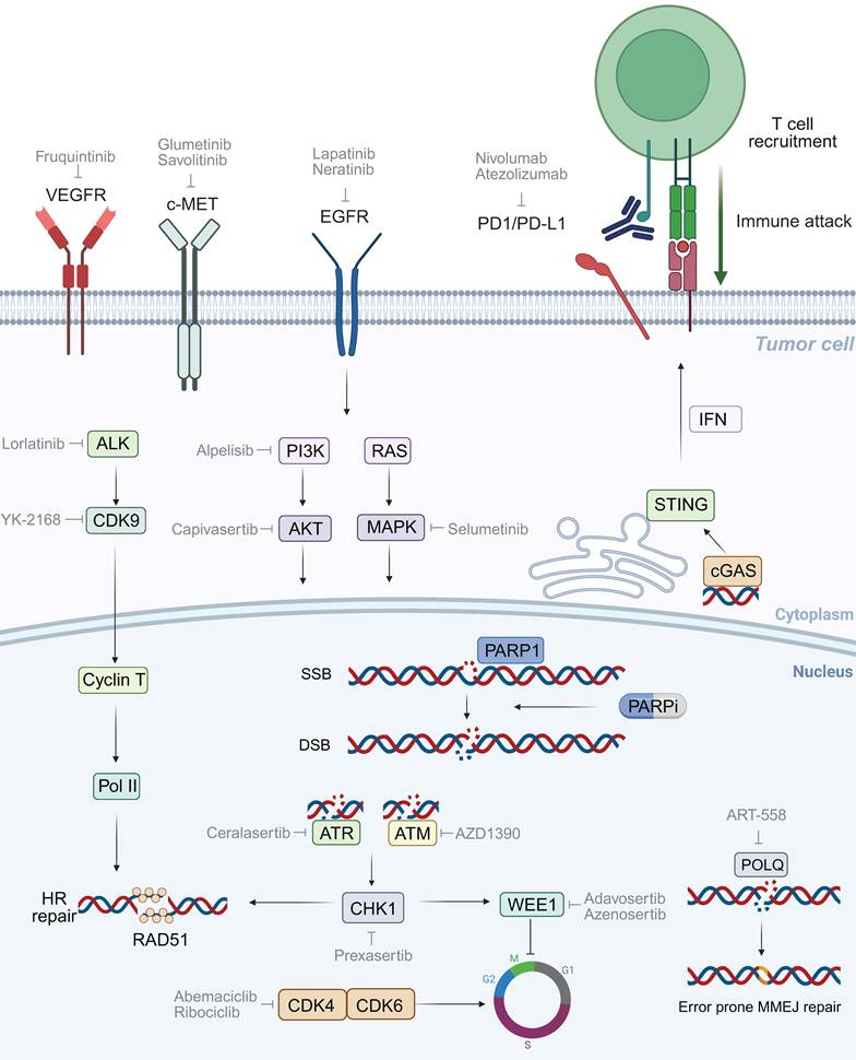
Therapeutic strategies should be interpreted according to both evidence level and treatment setting. A first-line combination may improve initial disease control without reversing an established resistance mechanism; a maintenance trial may enroll platinum-responsive, PARPi-naïve patients; and a preclinical resensitization experiment may demonstrate biological plausibility rather than clinical benefit. The most relevant evidence for overcoming acquired resistance comes from studies that explicitly enrolled patients progressing during or after prior PARPi. At progression, treatment decisions should remain disease-specific and consider platinum sensitivity, prior therapy, toxicity, pace of disease, and access to clinical trials. Biomarker-directed treatment is attractive, but there is limited prospective evidence that biomarker-matched interventions improve clinical outcomes. The sections below retain the original therapeutic categories while clearly distinguishing resistance-specific evidence from broader combination rationale (Figure 4).
Mechanisms of Dual-target inhibitors of PARP1. Investigational approaches include combinations involving immune checkpoint inhibitors, DNA-damage-response inhibitors, Polθ-directed agents, and selected kinase inhibitors. Evidence from approved indications or PARPi-naïve combination settings should not be interpreted as proof of reversal of established PARPi resistance; most strategies shown remain investigational in the resistance-specific setting.
5.1 Identifying Novel Predictive Biomarkers
Biomarker assessment should distinguish baseline prediction of PARPi benefit from reassessment after progression. At baseline, germline and tumor BRCA testing, biallelic status or loss of heterozygosity, tumor type, prior platinum sensitivity, and treatment setting remain central. After progression, the relevant question is which repair and survival functions are active at that time. Plasma ctDNA can be used for serial monitoring and may identify heterogeneous BRCA reversion clones, whereas tissue biopsy provides histologic context and enables RNA, methylation, protein, and functional assays. RAD51 nuclear foci offer a functional readout of HR competence and may distinguish PARPi-sensitive and PARPi-resistant tumors more directly than BRCA genotype alone [155, 156], but assay induction, cell-cycle controls, thresholds, and reproducibility require further standardization. No single test captures every phenotype: sequencing may detect reversion, methylation assays may identify promoter changes, RNA or protein analysis may support ABCB1-mediated efflux, and specialized pharmacodynamic assays may evaluate PAR metabolism. Together, these complementary assays can support reassessment and clinical-trial selection, but they should not be regarded as a validated prescriptive algorithm.
5.2 PARPi Combined with Radiotherapy
PARPi can radiosensitize tumor cells by limiting the repair of radiation-induced SSBs, increasing replication-associated lesions, and modifying stress-response pathways such as NF-κB [157]. PARP1/2-deficient cells are more sensitive to ionizing radiation, and low-dose olaparib has enhanced radiosensitization in HR-deficient experimental models [158]. Nevertheless, overlapping hematologic and tissue-specific toxicities may constrain dosing, and most studies have not been designed specifically for patients with established PARPi resistance. PARPi-radiotherapy combinations therefore remain investigational as strategies for overcoming resistance.
5.3 PARPi Combined with Immune Checkpoint Inhibitors
PARPi-induced DNA damage can activate innate immune signaling, providing a rationale for combination with immune checkpoint inhibitors (ICIs) targeting PD-1/PD-L1 or CTLA-4 [159]. Early studies have reported activity in several solid tumors. For example, olaparib plus durvalumab, with or without bevacizumab, produced high response rates in selected patients with platinum-sensitive recurrent ovarian cancer, particularly in germline BRCA-mutated disease [160]. However, many participants in such studies were not selected for established PARPi resistance, and results across ovarian cancer trials have been inconsistent. Rucaparib plus nivolumab did not consistently outperform PARPi alone, and benefit was not reliably predicted by HRD, BRCA status, or PD-L1 expression [161]. PARPi-ICI combinations should therefore not be described as proven resistance-reversal therapy. Better selection based on functional HR status, immune context, and prior PARPi exposure is required.
5.4 PARPi Combined with DNA Damage Response Inhibitors
ATR, ATM, CHK1, and WEE1 coordinate cell-cycle checkpoints and replication stress. Inhibiting these pathways can prevent arrest and repair, increase ssDNA and fork instability, reduce RAD51-mediated recovery, and force damaged cells into replication catastrophe or premature mitosis [162]. These effects provide a strong rationale for combination with PARPi, particularly when resistant tumors remain dependent on replication-stress checkpoints. Preclinical models and early clinical studies in PARPi-exposed ovarian cancer have shown signals of resensitization, including in BRCA2-mutated disease, but the available cohorts are generally small and heterogeneous. Accordingly, ATR/CHK1-directed combinations should be considered promising but investigational rather than established standards for PARPi resistance.
WEE1 regulates the G2/M checkpoint; its inhibition promotes mitotic entry before DNA repair is complete, leading to genomic instability and mitotic catastrophe [163, 164]. WEE1 inhibitors have shown activity in selected platinum-resistant ovarian cancers, including tumors with CCNE1 amplification or TP53 alterations, either alone or with chemotherapy [165, 166]. However, hematologic and gastrointestinal toxicities are substantial, particularly when WEE1 inhibition is combined with PARPi or cytotoxic agents. Current development therefore emphasizes dose optimization, intermittent or sequential schedules, and biomarker-enriched populations. WEE1-directed treatment remains investigational in PARPi-resistant disease.
Polθ, encoded by POLQ, is a key enzyme in theta-mediated end joining and is frequently required for the survival of HR-deficient cells. Genetic or pharmacologic Polθ inhibition can produce synthetic lethality and resensitize PARPi-resistant models [167-169]. Compounds such as novobiocin, ART558, and related agents have generated encouraging preclinical results, while clinical development remains early. No Polθ inhibitor has yet demonstrated efficacy in a prospectively defined PARPi-resistant population, and the relationship between POLQ dependence, BRCA reversion, and restored HR requires careful biomarker assessment.
5.5 PARPi Combined with Protein Kinase Inhibitors
Abnormal kinase signaling can influence HR-gene expression, replication stress, angiogenesis, and PARP1 activity. These observations have motivated combination strategies and dual-target molecules that inhibit PARP1 together with EGFR, CDK4/6, VEGFR, or other kinases [170, 171]. EGFR and downstream RAS-MAPK or PI3K-AKT signaling can alter proliferation and DNA repair dependence, while kinase-mediated phosphorylation of PARP1 may reduce inhibitor binding [172]. Antiangiogenic approaches may increase hypoxia and replication stress, and CDK4/6 inhibition can reduce the expression of HR proteins in some models, creating a transient HR-deficient phenotype [173]. These concepts are mechanistically attractive, but most dual PARP-kinase compounds and many broad kinase combinations remain preclinical or early-phase. Evidence from BRCA wild-type or PARPi-naïve models should not be interpreted as proof that these strategies reverse acquired resistance.
Receptor tyrosine kinases provide additional examples of context-dependent resistance biology. c-MET-mediated phosphorylation of PARP1 at Y907 can enhance PARP1 activity and reduce PARPi binding, and inhibition of c-MET or EGFR has restored sensitivity in experimental systems and selected tumor specimens [141]. ALK signaling may increase HR-gene transcription through CDK9 and has been linked to PARPi or platinum resistance. ALK inhibitors combined with olaparib have produced tumor regression in cell-line-derived and patient-derived xenograft models [174, 175]. These findings support clinical investigation but remain predominantly preclinical. Regulatory proteins such as HPF1, YB-1, Sam68, BANF1, and TRIP12 may also alter PARP1 activity, stability, or PARylation [33]. Their value as patient-selection biomarkers has not been established.
6. Conclusions and Perspectives
PARPi have transformed the treatment of selected BRCA1/2-mutated and HR-deficient cancers by exploiting synthetic lethality. Their cytotoxicity is now understood as a nonlinear interaction among catalytic inhibition, PARP1 trapping, post-replicative ssDNA gaps, replication stress, and transcription-replication conflicts rather than as a single SSB-to-DSB pathway. Resistance is similarly heterogeneous. BRCA1/2 reversion and restoration of HR have the strongest clinical support, whereas fork protection, altered PARP/PARG biology, and drug efflux are context-dependent and less readily measured in routine practice. Epigenetic and microenvironmental adaptations remain important research areas but are supported mainly by preclinical evidence. Future studies should combine serial ctDNA, tissue-based molecular and functional assays, and clearly defined clinical settings to distinguish baseline insensitivity from acquired resistance. Trials should also separate PARPi-naïve combinations from true resistance-specific interventions and prospectively test whether biomarker-matched treatment improves outcomes. Selective PARP1 inhibitors such as saruparib may improve the therapeutic index, but they remain investigational and have not yet been proven to overcome a defined resistance mechanism. A clinically useful framework will therefore require both mechanistic precision and evidence-qualified interpretation.
Acknowledgements
Funding Statement
This work was supported by the Medical Research Projects of Jiangsu Provincial Health Commission (Z2022028).
Data Sharing Statement
No data was used for the research described in the article.
Author Contributions
All authors significantly contributed to the reported work, encompassing conception, study design, execution, data acquisition, analysis, and interpretation; participated in drafting, revising, or critically reviewing the article; provided final approval for the version to be published; consented to the journal to which the article has been submitted; and accepted accountability for all aspects of the work.
Competing Interests
The authors have declared that no competing interest exists.
References
1. Anderson CJ, Talmane L, Luft J, Connelly J, Nicholson MD, Verburg JC. et al. Strand-resolved mutagenicity of DNA damage and repair. Nature. 2024;630:744-51
2. Alexandrov LB, Kim J, Haradhvala NJ, Huang MN, Tian Ng AW, Wu Y. et al. The repertoire of mutational signatures in human cancer. Nature. 2020;578:94-101
3. Chatterjee N, Walker GC. Mechanisms of DNA damage, repair, and mutagenesis. Environ Mol Mutagen. 2017;58:235-63
4. Vignard J, Mirey G, Salles B. Ionizing-radiation induced DNA double-strand breaks: a direct and indirect lighting up. Radiother Oncol. 2013;108:362-9
5. Rieckher M, Gallrein C, Alquezar-Artieda N, Bourached-Silva N, Vaddavalli PL, Mares D. et al. Distinct DNA repair mechanisms prevent formaldehyde toxicity during development, reproduction and aging. Nucleic Acids Res. 2024;52:8271-85
6. Ciccia A, Elledge SJ. The DNA damage response: making it safe to play with knives. Mol Cell. 2010;40:179-204
7. Ranjha L, Howard SM, Cejka P. Main steps in DNA double-strand break repair: an introduction to homologous recombination and related processes. Chromosoma. 2018;127:187-214
8. Scully R, Walter JC, Nussenzweig A. One-ended and two-ended breaks at nickase-broken replication forks. DNA Repair (Amst). 2024;144:103783
9. Bouwman P, Jonkers J. The effects of deregulated DNA damage signalling on cancer chemotherapy response and resistance. Nat Rev Cancer. 2012;12:587-98
10. Schreiber V, Dantzer F, Ame J-C, de Murcia G. Poly(ADP-ribose): novel functions for an old molecule. Nat Rev Mol Cell Biol. 2006;7:517-28
11. Jackson SP, Bartek J. The DNA-damage response in human biology and disease. Nature. 2009;461:1071-8
12. Coon EA, Benarroch EE. DNA damage response: Selected review and neurologic implications. Neurology. 2018;90:367-76
13. Chang HHY, Pannunzio NR, Adachi N, Lieber MR. Non-homologous DNA end joining and alternative pathways to double-strand break repair. Nat Rev Mol Cell Biol. 2017;18:495-506
14. Lieber MR, Karanjawala ZE. Ageing, repetitive genomes and DNA damage. Nat Rev Mol Cell Biol. 2004;5:69-75
15. Valerie K, Povirk LF. Regulation and mechanisms of mammalian double-strand break repair. Oncogene. 2003;22:5792-812
16. Riballo E, Kühne M, Rief N, Doherty A, Smith GC, Recio MJ. et al. A pathway of double-strand break rejoining dependent upon ATM, Artemis, and proteins locating to gamma-H2AX foci. Mol Cell. 2004;16:715-24
17. Lieber MR. The mechanism of double-strand DNA break repair by the nonhomologous DNA end-joining pathway. Annu Rev Biochem. 2010;79:181-211
18. Drew Y, Zenke FT, Curtin NJ. DNA damage response inhibitors in cancer therapy: lessons from the past, current status and future implications. Nat Rev Drug Discov. 2025;24:19-39
19. Wyatt DW, Feng W, Conlin MP, Yousefzadeh MJ, Roberts SA, Mieczkowski P. et al. Essential Roles for Polymerase θ-Mediated End Joining in the Repair of Chromosome Breaks. Mol Cell. 2016;63:662-73
20. Koole W, van Schendel R, Karambelas AE, van Heteren JT, Okihara KL, Tijsterman M. A Polymerase Theta-dependent repair pathway suppresses extensive genomic instability at endogenous G4 DNA sites. Nat Commun. 2014;5:3216
21. Mateos-Gomez PA, Gong F, Nair N, Miller KM, Lazzerini-Denchi E, Sfeir A. Mammalian polymerase θ promotes alternative NHEJ and suppresses recombination. Nature. 2015;518:254-7
22. Moynahan ME, Jasin M. Mitotic homologous recombination maintains genomic stability and suppresses tumorigenesis. Nat Rev Mol Cell Biol. 2010;11:196-207
23. Becker JR, Clifford G, Bonnet C, Groth A, Wilson MD, Chapman JR. BARD1 reads H2A lysine 15 ubiquitination to direct homologous recombination. Nature. 2021;596:433-7
24. Swift ML, Beishline K, Flashner S, Azizkhan-Clifford J. DSB repair pathway choice is regulated by recruitment of 53BP1 through cell cycle-dependent regulation of Sp1. Cell Rep. 2021;34:108840
25. Kijas AW, Lim YC, Bolderson E, Cerosaletti K, Gatei M, Jakob B. et al. ATM-dependent phosphorylation of MRE11 controls extent of resection during homology directed repair by signalling through Exonuclease 1. Nucleic Acids Res. 2015;43:8352-67
26. Shakya R, Reid LJ, Reczek CR, Cole F, Egli D, Lin CS. et al. BRCA1 tumor suppression depends on BRCT phosphoprotein binding, but not its E3 ligase activity. Science. 2011;334:525-8
27. Wong AK, Ormonde PA, Pero R, Chen Y, Lian L, Salada G. et al. Characterization of a carboxy-terminal BRCA1 interacting protein. Oncogene. 1998;17:2279-85
28. Aravind L, Koonin EV. Prokaryotic homologs of the eukaryotic DNA-end-binding protein Ku, novel domains in the Ku protein and prediction of a prokaryotic double-strand break repair system. Genome Res. 2001;11:1365-74
29. Szostak JW, Orr-Weaver TL, Rothstein RJ, Stahl FW. The double-strand-break repair model for recombination. Cell. 1983;33:25-35
30. Sale JE. Competition, collaboration and coordination-determining how cells bypass DNA damage. J Cell Sci. 2012;125:1633-43
31. Berti M, Cortez D, Lopes M. The plasticity of DNA replication forks in response to clinically relevant genotoxic stress. Nat Rev Mol Cell Biol. 2020;21:633-51
32. Xu Z, Xie H, Song L, Huang Y, Huang J. BRCA1 and BRCA2 in DNA damage and replication stress response: Insights into their functions, mechanisms, and implications for cancer treatment. DNA Repair (Amst). 2025;150:103847
33. Rose M, Burgess JT, O'Byrne K, Richard DJ, Bolderson E. PARP Inhibitors: Clinical Relevance, Mechanisms of Action and Tumor Resistance. Front Cell Dev Biol. 2020;8:564601
34. Bryant HE, Schultz N, Thomas HD, Parker KM, Flower D, Lopez E. et al. Specific killing of BRCA2-deficient tumours with inhibitors of poly(ADP-ribose) polymerase. Nature. 2005;434:913-7
35. Penson RT, Valencia RV, Cibula D, Colombo N, Leath CA 3rd, Bidziński M. et al. Olaparib Versus Nonplatinum Chemotherapy in Patients with Platinum-Sensitive Relapsed Ovarian Cancer and a Germline BRCA1/2 Mutation (SOLO3): A Randomized Phase III Trial. J Clin Oncol. 2020;38:1164-74
36. Monk BJ, Parkinson C, Lim MC, O'Malley DM, Oaknin A, Wilson MK. et al. A Randomized, Phase III Trial to Evaluate Rucaparib Monotherapy as Maintenance Treatment in Patients with Newly Diagnosed Ovarian Cancer (ATHENA-MONO/GOG-3020/ENGOT-ov45). J Clin Oncol. 2022;40:3952-64
37. Ray Chaudhuri A, Nussenzweig A. The multifaceted roles of PARP1 in DNA repair and chromatin remodelling. Nat Rev Mol Cell Biol. 2017;18:610-21
38. Murai J, Pommier Y. BRCAness, Homologous Recombination Deficiencies, and Synthetic Lethality. Cancer Res. 2023;83:1173-4
39. Turner N, Tutt A, Ashworth A. Hallmarks of 'BRCAness' in sporadic cancers. Nat Rev Cancer. 2004;4:814-9
40. Wang Y, Bernhardy AJ, Cruz C, Krais JJ, Nacson J, Nicolas E. et al. The BRCA1-Δ11q Alternative Splice Isoform Bypasses Germline Mutations and Promotes Therapeutic Resistance to PARP Inhibition and Cisplatin. Cancer Res. 2016;76:2778-90
41. Zhu H, Wei M, Xu J, Hua J, Liang C, Meng Q. et al. PARP inhibitors in pancreatic cancer: molecular mechanisms and clinical applications. Mol Cancer. 2020;19:49
42. Maxwell KN, Wubbenhorst B, Wenz BM, De Sloover D, Pluta J, Emery L. et al. BRCA locus-specific loss of heterozygosity in germline BRCA1 and BRCA2 carriers. Nat Commun. 2017;8:319
43. Farmer H, McCabe N, Lord CJ, Tutt ANJ, Johnson DA, Richardson TB. et al. Targeting the DNA repair defect in BRCA mutant cells as a therapeutic strategy. Nature. 2005;434:917-21
44. Ashworth A. A Synthetic Lethal Therapeutic Approach: Poly(ADP) Ribose Polymerase Inhibitors for the Treatment of Cancers Deficient in DNA Double-Strand Break Repair. J Clin Oncol. 2008;26:3785-90
45. Besse B, Pons-Tostivint E, Park K, Hartl S, Forde PM, Hochmair MJ. et al. Biomarker-directed targeted therapy plus durvalumab in advanced non-small-cell lung cancer: a phase 2 umbrella trial. Nat Med. 2024;30:716-29
46. Robson M, Im SA, Senkus E, Xu B, Domchek SM, Masuda N. et al. Olaparib for Metastatic Breast Cancer in Patients with a Germline BRCA Mutation. N Engl J Med. 2017;377:523-33
47. Noordermeer SM, van Attikum H. PARP Inhibitor Resistance: A Tug-of-War in BRCA-Mutated Cells. Trends Cell Biol. 2019;29:820-34
48. Miki Y, Swensen J, Shattuck-Eidens D, Futreal PA, Harshman K, Tavtigian S. et al. A strong candidate for the breast and ovarian cancer susceptibility gene BRCA1. Science. 1994;266:66-71
49. Venkitaraman AR. Cancer suppression by the chromosome custodians, BRCA1 and BRCA2. Science. 2014;343:1470-5
50. Yoshida K, Miki Y. Role of BRCA1 and BRCA2 as regulators of DNA repair, transcription, and cell cycle in response to DNA damage. Cancer Sci. 2004;95:866-71
51. Reid LJ, Shakya R, Modi AP, Lokshin M, Cheng JT, Jasin M. et al. E3 ligase activity of BRCA1 is not essential for mammalian cell viability or homology-directed repair of double-strand DNA breaks. Proc Natl Acad Sci U S A. 2008;105:20876-81
52. Wu LC, Wang ZW, Tsan JT, Spillman MA, Phung A, Xu XL. et al. Identification of a RING protein that can interact in vivo with the BRCA1 gene product. Nat Genet. 1996;14:430-40
53. Prakash R, Zhang Y, Feng W, Jasin M. Homologous recombination and human health: the roles of BRCA1, BRCA2, and associated proteins. Cold Spring Harb Perspect Biol. 2015;7:a016600
54. Cortez D, Wang Y, Qin J, Elledge SJ. Requirement of ATM-dependent phosphorylation of brca1 in the DNA damage response to double-strand breaks. Science. 1999;286:1162-6
55. Xia B, Dorsman JC, Ameziane N, de Vries Y, Rooimans MA, Sheng Q. et al. Fanconi anemia is associated with a defect in the BRCA2 partner PALB2. Nat Genet. 2007;39:159-61
56. Williams RS, Green R, Glover JN. Crystal structure of the BRCT repeat region from the breast cancer-associated protein BRCA1. Nat Struct Biol. 2001;8:838-42
57. Ceppi I, Howard SM, Kasaciunaite K, Pinto C, Anand R, Seidel R. et al. CtIP promotes the motor activity of DNA2 to accelerate long-range DNA end resection. Proc Natl Acad Sci U S A. 2020;117:8859-69
58. Yu X, Chini CC, He M, Mer G, Chen J. The BRCT domain is a phospho-protein binding domain. Science. 2003;302:639-42
59. Wang B, Matsuoka S, Ballif BA, Zhang D, Smogorzewska A, Gygi SP. et al. Abraxas and RAP80 form a BRCA1 protein complex required for the DNA damage response. Science. 2007;316:1194-8
60. Wooster R, Neuhausen SL, Mangion J, Quirk Y, Ford D, Collins N. et al. Localization of a breast cancer susceptibility gene, BRCA2, to chromosome 13q12-13. Science. 1994;265:2088-90
61. Bignell G, Micklem G, Stratton MR, Ashworth A, Wooster R. The BRC repeats are conserved in mammalian BRCA2 proteins. Hum Mol Genet. 1997;6:53-8
62. Carreira A, Hilario J, Amitani I, Baskin RJ, Shivji MK, Venkitaraman AR. et al. The BRC repeats of BRCA2 modulate the DNA-binding selectivity of RAD51. Cell. 2009;136:1032-43
63. Jensen RB, Carreira A, Kowalczykowski SC. Purified human BRCA2 stimulates RAD51-mediated recombination. Nature. 2010;467:678-83
64. Zhang F, Ma J, Wu J, Ye L, Cai H, Xia B. et al. PALB2 Links BRCA1 and BRCA2 in the DNA-Damage Response. Curr Biol. 2009;19:524-9
65. Esashi F, Galkin VE, Yu X, Egelman EH, West SC. Stabilization of RAD51 nucleoprotein filaments by the C-terminal region of BRCA2. Nat Struct Mol Biol. 2007;14:468-74
66. Esashi F, Christ N, Gannon J, Liu Y, Hunt T, Jasin M. et al. CDK-dependent phosphorylation of BRCA2 as a regulatory mechanism for recombinational repair. Nature. 2005;434:598-604
67. Mishra AP, Hartford SA, Sahu S, Klarmann K, Chittela RK, Biswas K. et al. BRCA2-DSS1 interaction is dispensable for RAD51 recruitment at replication-induced and meiotic DNA double strand breaks. Nat Commun. 2022;13:1751
68. Yang H, Jeffrey PD, Miller J, Kinnucan E, Sun Y, Thoma NH. et al. BRCA2 function in DNA binding and recombination from a BRCA2-DSS1-ssDNA structure. Science. 2002;297:1837-48
69. Huang Y, Li W, Foo T, Ji JH, Wu B, Tomimatsu N. et al. DSS1 restrains BRCA2's engagement with dsDNA for homologous recombination, replication fork protection, and R-loop homeostasis. Nat Commun. 2024;15:7081
70. Kwon Y, Rösner H, Zhao W, Selemenakis P, He Z, Kawale AS. et al. DNA binding and RAD51 engagement by the BRCA2 C-terminus orchestrate DNA repair and replication fork preservation. Nat Commun. 2023;14:432
71. Yang H, Li Q, Fan J, Holloman WK, Pavletich NP. The BRCA2 homologue Brh2 nucleates RAD51 filament formation at a dsDNA-ssDNA junction. Nature. 2005;433:653-7
72. Patel KJ. Fanconi anemia and breast cancer susceptibility. Nat Genet. 2007;39:142-3
73. Daniels MJ, Wang Y, Lee M, Venkitaraman AR. Abnormal cytokinesis in cells deficient in the breast cancer susceptibility protein BRCA2. Science. 2004;306:876-9
74. Chen CC, Feng W, Lim PX, Kass EM, Jasin M. Homology-Directed Repair and the Role of BRCA1, BRCA2, and Related Proteins in Genome Integrity and Cancer. Annu Rev Cancer Biol. 2018;2:313-36
75. Anantha RW, Simhadri S, Foo TK, Miao S, Liu J, Shen Z. et al. Functional and mutational landscapes of BRCA1 for homology-directed repair and therapy resistance. Elife. 2017;6:e21350
76. Moynahan ME. The cancer connection: BRCA1 and BRCA2 tumor suppression in mice and humans. Oncogene. 2002;21:8994-9007
77. Tarsounas M, Sung P. The antitumorigenic roles of BRCA1-BARD1 in DNA repair and replication. Nat Rev Mol Cell Biol. 2020;21:284-99
78. Roy R, Chun J, Powell SN. BRCA1 and BRCA2: different roles in a common pathway of genome protection. Nat Rev Cancer. 2011;12:68-78
79. Escribano-Díaz C, Orthwein A, Fradet-Turcotte A, Xing M, Young Jordan TF, Tkáč J. et al. A Cell Cycle-Dependent Regulatory Circuit Composed of 53BP1-RIF1 and BRCA1-CtIP Controls DNA Repair Pathway Choice. Mol Cell. 2013;49:872-83
80. Gudmundsdottir K, Lord CJ, Witt E, Tutt AN, Ashworth A. DSS1 is required for RAD51 focus formation and genomic stability in mammalian cells. EMBO Rep. 2004;5:989-93
81. Foo TK, Xia B. BRCA1-Dependent and Independent Recruitment of PALB2-BRCA2-RAD51 in the DNA Damage Response and Cancer. Cancer Res. 2022;82:3191-7
82. Zhang F, Fan Q, Ren K, Andreassen PR. PALB2 functionally connects the breast cancer susceptibility proteins BRCA1 and BRCA2. Mol Cancer Res. 2009;7:1110-8
83. Lei T, Du S, Peng Z, Chen L. Multifaceted regulation and functions of 53BP1 in NHEJ-mediated DSB repair (Review). Int J Mol Med. 2022;50:90
84. Zhong Q, Xiao X, Qiu Y, Xu Z, Chen C, Chong B. et al. Protein posttranslational modifications in health and diseases: Functions, regulatory mechanisms, and therapeutic implications. MedComm (2020). 2023;4:e261
85. Wei H, Yu X. Functions of PARylation in DNA Damage Repair Pathways. Genomics Proteomics Bioinformatics. 2016;14:131-9
86. Satoh MS, Lindahl T. Role of poly(ADP-ribose) formation in DNA repair. Nature. 1992;356:356-8
87. Vyas S, Matic I, Uchima L, Rood J, Zaja R, Hay RT. et al. Family-wide analysis of poly(ADP-ribose) polymerase activity. Nat Commun. 2014;5:4426
88. Ke Y, Wang C, Zhang J, Zhong X, Wang R, Zeng X. et al. The Role of PARPs in Inflammation-and Metabolic-Related Diseases: Molecular Mechanisms and Beyond. Cells. 2019;8:1047
89. Ménissier de Murcia J, Ricoul M, Tartier L, Niedergang C, Huber A, Dantzer F. et al. Functional interaction between PARP-1 and PARP-2 in chromosome stability and embryonic development in mouse. EMBO J. 2003;22:2255-63
90. Martin-Hernandez K, Rodriguez-Vargas JM, Schreiber V, Dantzer F. Expanding functions of ADP-ribosylation in the maintenance of genome integrity. Semin Cell Dev Biol. 2017;63:92-101
91. Eustermann S, Wu WF, Langelier MF, Yang JC, Easton LE, Riccio AA. et al. Structural Basis of Detection and Signaling of DNA Single-Strand Breaks by Human PARP-1. Mol Cell. 2015;60:742-54
92. Huambachano O, Herrera F, Rancourt A, Satoh MS. Double-stranded DNA Binding Domain of Poly(ADP-ribose) Polymerase-1 and Molecular Insight into the Regulation of Its Activity. J Biol Chem. 2011;286:7149-60
93. Langelier MF, Pascal JM. PARP-1 mechanism for coupling DNA damage detection to poly(ADP-ribose) synthesis. Curr Opin Struct Biol. 2013;23:134-43
94. Hassler M, Ladurner AG. Towards a structural understanding of PARP1 activation and related signalling ADP-ribosyl-transferases. Curr Opin Struct Biol. 2012;22:721-9
95. Langelier MF, Planck JL, Roy S, Pascal JM. Structural basis for DNA damage-dependent poly(ADP-ribosyl)ation by human PARP-1. Science. 2012;336:728-32
96. Tulin A, Spradling A. Chromatin loosening by poly (ADP)-ribose polymerase (PARP) at Drosophila puff loci. Science. 2003;299:560-2
97. Gottschalk AJ, Timinszky G, Kong SE, Jin J, Cai Y, Swanson SK. et al. Poly(ADP-ribosyl)ation directs recruitment and activation of an ATP-dependent chromatin remodeler. Proc Natl Acad Sci U S A. 2009;106:13770-4
98. Langelier MF, Eisemann T, Riccio AA, Pascal JM. PARP family enzymes: regulation and catalysis of the poly(ADP-ribose) posttranslational modification. Curr Opin Struct Biol. 2018;53:187-98
99. Gagné JP, Isabelle M, Lo KS, Bourassa S, Hendzel MJ, Dawson VL. et al. Proteome-wide identification of poly(ADP-ribose) binding proteins and poly(ADP-ribose)-associated protein complexes. Nucleic Acids Res. 2008;36:6959-76
100. Jungmichel S, Rosenthal F, Altmeyer M, Lukas J, Hottiger MO, Nielsen ML. Proteome-wide identification of poly(ADP-Ribosyl)ation targets in different genotoxic stress responses. Mol Cell. 2013;52:272-85
101. Barkauskaite E, Jankevicius G, Ahel I. Structures and Mechanisms of Enzymes Employed in the Synthesis and Degradation of PARP-Dependent Protein ADP-Ribosylation. Mol Cell. 2015;58:935-46
102. Groslambert J, Prokhorova E, Wondisford AR, Tromans-Coia C, Giansanti C, Jansen J. et al. The interplay of TARG1 and PARG protects against genomic instability. Cell Rep. 2023;42:113113
103. Li Y, Liu CF, Rao GW. A Review on Poly (ADP-ribose) Polymerase (PARP) Inhibitors and Synthetic Methodologies. Curr Med Chem. 2021;28:1565-84
104. Andronikou C, Rottenberg S. Studying PAR-Dependent Chromatin Remodeling to Tackle PARPi Resistance. Trends Mol Med. 2021;27:630-42
105. Tew WP, Lacchetti C, Ellis A, Maxian K, Banerjee S, Bookman M. et al. PARP Inhibitors in the Management of Ovarian Cancer: ASCO Guideline. J Clin Oncol. 2020;38:3468-93
106. Petropoulos M, Karamichali A, Rossetti GG, Freudenmann A, Iacovino LG, Dionellis VS. et al. Transcription-replication conflicts underlie sensitivity to PARP inhibitors. Nature. 2024;628:433-41
107. Soung YH, Chung J. Combination Treatment Strategies to Overcome PARP Inhibitor Resistance. Biomolecules. 2023;13:1480
108. Kim G, Ison G, McKee AE, Zhang H, Tang S, Gwise T. et al. FDA Approval Summary: Olaparib Monotherapy in Patients with Deleterious Germline BRCA-Mutated Advanced Ovarian Cancer Treated with Three or More Lines of Chemotherapy. Clin Cancer Res. 2015;21:4257-61
109. Banerjee S, Moore KN, Colombo N, Scambia G, Kim BG, Oaknin A. et al. Maintenance olaparib for patients with newly diagnosed advanced ovarian cancer and a BRCA mutation (SOLO1/GOG 3004): 5-year follow-up of a randomised, double-blind, placebo-controlled, phase 3 trial. Lancet Oncol. 2021;22:1721-31
110. Mateo J, Porta N, Bianchini D, McGovern U, Elliott T, Jones R. et al. Olaparib in patients with metastatic castration-resistant prostate cancer with DNA repair gene aberrations (TOPARP-B): a multicentre, open-label, randomised, phase 2 trial. Lancet Oncol. 2020;21:162-74
111. Anscher MS, Chang E, Gao X, Gong Y, Weinstock C, Bloomquist E. et al. FDA Approval Summary: Rucaparib for the Treatment of Patients with Deleterious BRCA-Mutated Metastatic Castrate-Resistant Prostate Cancer. Oncologist. 2021;26:139-46
112. Ison G, Howie LJ, Amiri-Kordestani L, Zhang L, Tang S, Sridhara R. et al. FDA Approval Summary: Niraparib for the Maintenance Treatment of Patients with Recurrent Ovarian Cancer in Response to Platinum-Based Chemotherapy. Clin Cancer Res. 2018;24:4066-71
113. de Bono J, Ramanathan RK, Mina L, Chugh R, Glaspy J, Rafii S. et al. Phase I, Dose-Escalation, Two-Part Trial of the PARP Inhibitor Talazoparib in Patients with Advanced Germline BRCA1/2 Mutations and Selected Sporadic Cancers. Cancer Discov. 2017;7:620-9
114. Lee A. Fuzuloparib: First Approval. Drugs. 2021;81:1221-6
115. Chu YY, Yam C, Yamaguchi H, Hung MC. Biomarkers beyond BRCA: promising combinatorial treatment strategies in overcoming resistance to PARP inhibitors. J Biomed Sci. 2022;29:86
116. Johannes JW, Balazs A, Barratt D, Bista M, Chuba MD, Cosulich S. et al. Discovery of 5-{4-[(7-Ethyl-6-oxo-5,6-dihydro-1,5-naphthyridin-3-yl)methyl]piperazin-1-yl}-N-methylpyridine-2-carboxamide (AZD5305): A PARP1-DNA Trapper with High Selectivity for PARP1 over PARP2 and Other PARPs. J Med Chem. 2021;64:14498-512
117. Dias MP, Moser SC, Ganesan S, Jonkers J. Understanding and overcoming resistance to PARP inhibitors in cancer therapy. Nat Rev Clin Oncol. 2021;18:773-91
118. McGranahan N, Swanton C. Clonal Heterogeneity and Tumor Evolution: Past, Present, and the Future. Cell. 2017;168:613-28
119. Prager BC, Xie Q, Bao S, Rich JN. Cancer Stem Cells: The Architects of the Tumor Ecosystem. Cell Stem Cell. 2019;24:41-53
120. Piombino C, Pipitone S, Tonni E, Mastrodomenico L, Oltrecolli M, Tchawa C. et al. Homologous Recombination Repair Deficiency in Metastatic Prostate Cancer: New Therapeutic Opportunities. Int J Mol Sci. 2024;25:4624
121. Goodall J, Mateo J, Yuan W, Mossop H, Porta N, Miranda S. et al. Circulating Cell-Free DNA to Guide Prostate Cancer Treatment with PARP Inhibition. Cancer Discov. 2017;7:1006-17
122. Walmsley CS, Jonsson P, Cheng ML, McBride S, Kaeser C, Vargas HA. et al. Convergent evolution of BRCA2 reversion mutations under therapeutic pressure by PARP inhibition and platinum chemotherapy. NPJ Precis Oncol. 2024;8:34
123. Polkinghorn WR, Parker JS, Lee MX, Kass EM, Spratt DE, Iaquinta PJ. et al. Androgen Receptor Signaling Regulates DNA Repair in Prostate Cancers. Cancer Discov. 2013;3:1245-53
124. Schiewer MJ, Goodwin JF, Han S, Brenner JC, Augello MA, Dean JL. et al. Dual Roles of PARP-1 Promote Cancer Growth and Progression. Cancer Discov. 2012;2:1134-49
125. Johnson N, Johnson SF, Yao W, Li YC, Choi YE, Bernhardy AJ. et al. Stabilization of mutant BRCA1 protein confers PARP inhibitor and platinum resistance. Proc Natl Acad Sci U S A. 2013;110:17041-6
126. Tao H, Liu S, Huang D, Han X, Wu X, Shao YW. et al. Acquired multiple secondary BRCA2 mutations upon PARPi resistance in a metastatic pancreatic cancer patient harboring a BRCA2 germline mutation. Am J Transl Res. 2020;12:612-7
127. Marzio A, Puccini J, Kwon Y, Maverakis NK, Arbini A, Sung P. et al. The F-Box Domain-Dependent Activity of EMI1 Regulates PARPi Sensitivity in Triple-Negative Breast Cancers. Mol Cell. 2019;73:224-37.e6
128. Ronson GE, Piberger AL, Higgs MR, Olsen AL, Stewart GS, McHugh PJ. et al. PARP1 and PARP2 stabilise replication forks at base excision repair intermediates through Fbh1-dependent Rad51 regulation. Nat Commun. 2018;9:746
129. Gupta R, Somyajit K, Narita T, Maskey E, Stanlie A, Kremer M. et al. DNA Repair Network Analysis Reveals Shieldin as a Key Regulator of NHEJ and PARP Inhibitor Sensitivity. Cell. 2018;173:972-88.e23
130. Bunting SF, Callén E, Wong N, Chen HT, Polato F, Gunn A. et al. 53BP1 inhibits homologous recombination in Brca1-deficient cells by blocking resection of DNA breaks. Cell. 2010;141:243-54
131. Xu G, Chapman JR, Brandsma I, Yuan J, Mistrik M, Bouwman P. et al. REV7 counteracts DNA double-strand break resection and affects PARP inhibition. Nature. 2015;521:541-4
132. Choi YE, Meghani K, Brault ME, Leclerc L, He YJ, Day TA. et al. Platinum and PARP Inhibitor Resistance Due to Overexpression of MicroRNA-622 in BRCA1-Mutant Ovarian Cancer. Cell Rep. 2016;14:429-39
133. Lin KK, Harrell MI, Oza AM, Oaknin A, Ray-Coquard I, Tinker AV. et al. BRCA Reversion Mutations in Circulating Tumor DNA Predict Primary and Acquired Resistance to the PARP Inhibitor Rucaparib in High-Grade Ovarian Carcinoma. Cancer Discov. 2019;9:210-9
134. Lemaçon D, Jackson J, Quinet A, Brickner JR, Li S, Yazinski S. et al. MRE11 and EXO1 nucleases degrade reversed forks and elicit MUS81-dependent fork rescue in BRCA2-deficient cells. Nat Commun. 2017;8:860
135. Kolinjivadi AM, Sannino V, De Antoni A, Zadorozhny K, Kilkenny M, Técher H. et al. Smarcal1-Mediated Fork Reversal Triggers Mre11-Dependent Degradation of Nascent DNA in the Absence of Brca2 and Stable Rad51 Nucleofilaments. Mol Cell. 2017;67:867-81.e7
136. Taglialatela A, Alvarez S, Leuzzi G, Sannino V, Ranjha L, Huang JW. et al. Restoration of Replication Fork Stability in BRCA1- and BRCA2-Deficient Cells by Inactivation of SNF2-Family Fork Remodelers. Mol Cell. 2017;68:414-30.e8
137. Rondinelli B, Gogola E, Yücel H, Duarte AA, van de Ven M, van der Sluijs R. et al. EZH2 promotes degradation of stalled replication forks by recruiting MUS81 through histone H3 trimethylation. Nat Cell Biol. 2017;19:1371-8
138. Dungrawala H, Bhat KP, Le Meur R, Chazin WJ, Ding X, Sharan SK. et al. RADX Promotes Genome Stability and Modulates Chemosensitivity by Regulating RAD51 at Replication Forks. Mol Cell. 2017;67:374-86.e5
139. Bossennec M, Di Roio A, Caux C, Ménétrier-Caux C. MDR1 in immunity: friend or foe? Oncoimmunology. 2018;7:e1499388
140. Pettitt SJ, Krastev DB, Brandsma I, Dréan A, Song F, Aleksandrov R. et al. Genome-wide and high-density CRISPR-Cas9 screens identify point mutations in PARP1 causing PARP inhibitor resistance. Nat Commun. 2018;9:1849
141. Du Y, Yamaguchi H, Wei Y, Hsu JL, Wang HL, Hsu YH. et al. Blocking c-Met-mediated PARP1 phosphorylation enhances anti-tumor effects of PARP inhibitors. Nat Med. 2016;22:194-201
142. Nespolo A, Stefenatti L, Pellarin I, Gambelli A, Rampioni Vinciguerra GL, Karimbayli J. et al. USP1 deubiquitinates PARP1 to regulate its trapping and PARylation activity. Sci Adv. 2024;10:eadp6567
143. Zhan Z, Zhang J, Liang H, Wang C, Hong L, Liu W. KAT6A Condensates Impair PARP1 Trapping of PARP Inhibitors in Ovarian Cancer. Adv Sci (Weinh). 2024;11:e2400140
144. Ma H, Qi G, Han F, Lu W, Peng J, Li R. et al. HMGB3 promotes PARP inhibitor resistance through interacting with PARP1 in ovarian cancer. Cell Death Dis. 2022;13:263
145. Gogola E, Duarte AA, de Ruiter JR, Wiegant WW, Schmid JA, de Bruijn R. et al. Selective Loss of PARG Restores PARylation and Counteracts PARP Inhibitor-Mediated Synthetic Lethality. Cancer Cell. 2018;33:1078-93.e12
146. Houl JH, Ye Z, Brosey CA, Balapiti-Modarage LPF, Namjoshi S, Bacolla A. et al. Selective small molecule PARG inhibitor causes replication fork stalling and cancer cell death. Nat Commun. 2019;10:5654
147. Yi X-F, Gao R-L, Sun L, Wu Z-X, Zhang S-L, Huang L-T. et al. Dual antitumor immunomodulatory effects of PARP inhibitor on the tumor microenvironment: A counterbalance between anti-tumor and pro-tumor. Biomed Pharmacother. 2023;163:114770
148. Qiu J, Ren T, Liu Q, Jiang Q, Wu T, Cheng LC. et al. Dissecting the Distinct Tumor Microenvironments of HRD and HRP Ovarian Cancer: Implications for Targeted Therapies to Overcome PARPi Resistance in HRD Tumors and Refractoriness in HRP Tumors. Adv Sci (Weinh). 2024;11:2309755
149. Shen J, Zhao W, Ju Z, Wang L, Peng Y, Labrie M. et al. PARPi Triggers the STING-Dependent Immune Response and Enhances the Therapeutic Efficacy of Immune Checkpoint Blockade Independent of BRCAness. Cancer Res. 2019;79:311-9
150. Xia Y, Huang P, Qian YY, Wang Z, Jin N, Li X. et al. PARP inhibitors enhance antitumor immune responses by triggering pyroptosis via TNF-caspase 8-GSDMD/E axis in ovarian cancer. J Immunother Cancer. 2024;12:e009032
151. Lu Y, Fu W, Xing W, Wu H, Zhang C, Xu D. Transcriptional regulation mechanism of PARP1 and its application in disease treatment. Epigenetics Chromatin. 2024;17:26
152. Beneyton A, Nonfoux L, Gagné JP, Rodrigue A, Kothari C, Atalay N. et al. The dynamic process of covalent and non-covalent PARylation in the maintenance of genome integrity: a focus on PARP inhibitors. NAR Cancer. 2023;5:zcad043
153. Tu Z, Wu L, Luo H, Li J, Lv S, Ye M. et al. Systematic and Multi-Omics Prognostic Analysis of Lysine Acetylation Regulators in Glioma. Front Mol Biosci. 2021;8:587516
154. Liu WW, Zhang ZY, Wang F, Wang H. Emerging roles of m6A RNA modification in cancer therapeutic resistance. Exp Hematol Oncol. 2023;12:21
155. Heeke AL, Pishvaian MJ, Lynce F, Xiu J, Brody JR, Chen WJ. et al. Prevalence of Homologous Recombination-Related Gene Mutations Across Multiple Cancer Types. JCO Precis Oncol. 2018;2:PO.17.00286
156. Pellegrino B, Herencia-Ropero A, Llop-Guevara A, Pedretti F, Moles-Fernández A, Viaplana C. et al. Preclinical In vivo Validation of the RAD51 Test for Identification of Homologous Recombination-Deficient Tumors and Patient Stratification. Cancer Res. 2022;82:1646-57
157. Sharma RA, Plummer R, Stock JK, Greenhalgh TA, Ataman O, Kelly S. et al. Clinical development of new drug-radiotherapy combinations. Nat Rev Clin Oncol. 2016;13:627-42
158. Mao Y, Huang X, Shuang Z, Lin G, Wang J, Duan F. et al. PARP inhibitor olaparib sensitizes cholangiocarcinoma cells to radiation. Cancer Med. 2018;7:1285-96
159. Miao H, Fang Y, Pan C, Yang H, Wang Z, Qi Y. et al. Transforming the landscape of cancer treatment with seven promising novel therapies: evolution and future perspectives. Medicine Plus. 2025;2:100087
160. Domchek SM, Postel-Vinay S, Im SA, Park YH, Delord JP, Italiano A. et al. Olaparib and durvalumab in patients with germline BRCA-mutated metastatic breast cancer (MEDIOLA): an open-label, multicentre, phase 1/2, basket study. Lancet Oncol. 2020;21:1155-64
161. Liu JF, Gaillard S, Wahner Hendrickson AE, Yeku O, Diver E, Gunderson Jackson C. et al. Niraparib, Dostarlimab, and Bevacizumab as Combination Therapy in Pretreated, Advanced Platinum-Resistant Ovarian Cancer: Findings from Cohort A of the OPAL Phase II Trial. JCO Precis Oncol. 2024;8:e2300693
162. Riches LC, Trinidad AG, Hughes G, Jones GN, Hughes AM, Thomason AG. et al. Pharmacology of the ATM Inhibitor AZD0156: Potentiation of Irradiation and Olaparib Responses Preclinically. Mol Cancer Ther. 2020;19:13-25
163. Ronco C, Martin AR, Demange L, Benhida R. ATM, ATR, CHK1, CHK2 and WEE1 inhibitors in cancer and cancer stem cells. Medchemcomm. 2017;8:295-319
164. Oza AM, Cibula D, Benzaquen AO, Poole C, Mathijssen RH, Sonke GS. et al. Olaparib combined with chemotherapy for recurrent platinum-sensitive ovarian cancer: a randomised phase 2 trial. Lancet Oncol. 2015;16:87-97
165. Blandino G, Di Agostino S. New therapeutic strategies to treat human cancers expressing mutant p53 proteins. J Exp Clin Cancer Res. 2018;37:30
166. Hu Y, Guo M. Synthetic lethality strategies: Beyond BRCA1/2 mutations in pancreatic cancer. Cancer Sci. 2020;111:3111-21
167. Zatreanu D, Robinson HMR, Alkhatib O, Boursier M, Finch H, Geo L. et al. Polθ inhibitors elicit BRCA-gene synthetic lethality and target PARP inhibitor resistance. Nat Commun. 2021;12:3636
168. Bubenik M, Mader P, Mochirian P, Vallée F, Clark J, Truchon JF. et al. Identification of RP-6685, an Orally Bioavailable Compound that Inhibits the DNA Polymerase Activity of Polθ. J Med Chem. 2022;65:13198-215
169. Zhou J, Gelot C, Pantelidou C, Li A, Yücel H, Davis RE. et al. A first-in-class Polymerase Theta Inhibitor selectively targets Homologous-Recombination-Deficient Tumors. Nat Cancer. 2021;2:598-610
170. Huang R, Zhou PK. DNA damage repair: historical perspectives, mechanistic pathways and clinical translation for targeted cancer therapy. Signal Transduct Target Ther. 2021;6:254
171. Goncalves A, Mezni E, Bertucci F. Combining poly(ADP-ribose) polymerase inhibitors and immune checkpoint inhibitors in breast cancer: rationale and preliminary clinical results. Curr Opin Oncol. 2020;32:585-93
172. Abbotts R, Dellomo AJ, Rassool FV. Pharmacologic Induction of BRCAness in BRCA-Proficient Cancers: Expanding PARP Inhibitor Use. Cancers (Basel). 2022;14:2640
173. Goel S, DeCristo MJ, Watt AC, BrinJones H, Sceneay J, Li BB. et al. CDK4/6 inhibition triggers anti-tumour immunity. Nature. 2017;548:471-5
174. Chu YY, Chen MK, Wei Y, Lee HH, Xia W, Wang YN. et al. Targeting the ALK-CDK9-Tyr19 kinase cascade sensitizes ovarian and breast tumors to PARP inhibition via destabilization of the P-TEFb complex. Nat Cancer. 2022;3:1211-27
175. Santiago-O'Farrill JM, Blessing Bollu A, Yang H, Orellana V, Pina M, Zhang X. et al. Crizotinib Enhances PARP Inhibitor Efficacy in Ovarian Cancer Cells and Xenograft Models by Inducing Autophagy. Mol Cancer Res. 2024;22:840-51
176. Ismail T, Alzneika S, Riguene E, Al-maraghi S, Alabdulrazzak A, Al-Khal N. et al. BRCA1 and Its Vulnerable C-Terminal BRCT Domain: Structure, Function, Genetic Mutations and Links to Diagnosis and Treatment of Breast and Ovarian Cancer. Pharmaceuticals. 2024;17:333
177. Millot GA, Carvalho MA, Caputo SM, Vreeswijk MP, Brown MA, Webb M. et al. A guide for functional analysis of BRCA1 variants of uncertain significance. Hum Mutat. 2012;33:1526-37
178. Chen C-F, Li S, Chen Y, Chen P-L, Sharp ZD, Lee W-H. The Nuclear Localization Sequences of the BRCA1 Protein Interact with the Importin-α Subunit of the Nuclear Transport Signal Receptor. J Biol Chem. 1996;271:32863-8
179. Yu X, Chini CCS, He M, Mer G, Chen J. The BRCT Domain Is a Phospho-Protein Binding Domain. Science. 2003;302:639-42
180. Le HP, Heyer WD, Liu J. Guardians of the Genome: BRCA2 and Its Partners. Genes (Basel). 2021;12:1229
181. Chatterjee G, Jimenez-Sainz J, Presti T, Nguyen T, Jensen RB. Distinct binding of BRCA2 BRC repeats to RAD51 generates differential DNA damage sensitivity. Nucleic Acids Res. 2016;44:5256-70
182. Andreassen PR, Seo J, Wiek C, Hanenberg H. Understanding BRCA2 Function as a Tumor Suppressor Based on Domain-Specific Activities in DNA Damage Responses. Genes (Basel). 2021;12:1034
183. Chambon P, Weill JD, Mandel P. Nicotinamide mononucleotide activation of a new DNA-dependent polyadenylic acid synthesizing nuclear enzyme. Biochem Biophys Res Commun. 1963;11:39-43
184. Yamada M, Miwa M, Sugimura T. Studies on poly (adenosine diphosphate-ribose). X. Properties of a partially purified poly (adenosine diphosphate-ribose) polymerase. Arch Biochem Biophys. 1971;146:579-86
185. Poirier GG, de Murcia G, Jongstra-Bilen J, Niedergang C, Mandel P. Poly(ADP-ribosyl)ation of polynucleosomes causes relaxation of chromatin structure. Proc Natl Acad Sci U S A. 1982;79:3423-7
186. Benjamin RC, Gill DM. ADP-ribosylation in mammalian cell ghosts. Dependence of poly(ADP-ribose) synthesis on strand breakage in DNA. J Biol Chem. 1980;255:10493-501
187. Amé JC, Rolli V, Schreiber V, Niedergang C, Apiou F, Decker P. et al. PARP-2, A novel mammalian DNA damage-dependent poly(ADP-ribose) polymerase. J Biol Chem. 1999;274:17860-8
188. Hall JM, Lee MK, Newman B, Morrow JE, Anderson LA, Huey B. et al. Linkage of Early-Onset Familial Breast Cancer to Chromosome 17q21. Science. 1990;250:1684-9
189. Fong PC, Boss DS, Yap TA, Tutt A, Wu P, Mergui-Roelvink M. et al. Inhibition of Poly(ADP-Ribose) Polymerase in Tumors from BRCA Mutation Carriers. N Engl J Med. 2009;361:123-34
190. Kolesnichenko M, Scheidereit C. Synthetic lethality by PARP inhibitors: new mechanism uncovered based on unresolved transcription-replication conflicts. Signal Transduct Target Ther. 2024;9:179
Author contact
Corresponding author: yztalijiecom (Jie Li); jzt880521com (Zhitao Jiang)