Impact Factor ISSN: 1837-9664




J Cancer 2026; 17(8):1531-1545. doi:10.7150/jca.135267 This issue Cite
Research Paper
Mutation Patterns Define Clinical Heterogeneity in Acute Myeloid Leukemia
Zhengrong Xu1,3#, Ying Zheng1,3#, Yi Zheng1,3#, Yanyan Yao1,3, Haili Geng1, Xiaofan Li1, Shao-yuan Wang1  , Lili Pan1,2,3
1. Fujian Institute of Hematology, Fujian Provincial Key Laboratory on Hematology, Department of Hematology, Fujian Medical University Union Hospital, Fuzhou 350001, PR China.
2. Translational Medicine Center on Hematology, Fujian Medical University, Fuzhou 350001, PR China.
3. Union Clinical Medical Colleges, Fujian Medical University, Fuzhou 350001, PR China.
# Zhengrong Xu, Ying Zheng, Yi Zheng contributed equally to this work.
Received 2026-3-28; Accepted 2026-7-25; Published 2026-8-12
Abstract
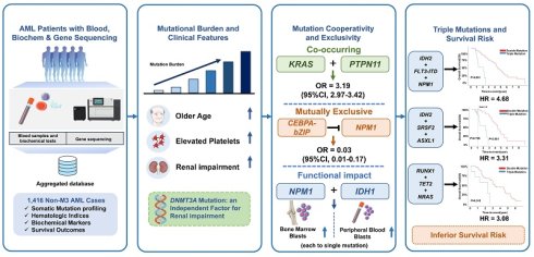
Background: Acute myeloid leukemia (AML) is a molecularly heterogeneous malignancy where next-generation sequencing (NGS) has revolutionized risk stratification and treatment paradigms. However, the interplay between mutation cooperativity, clinical phenotypes, and biochemical markers of organ dysfunction remains poorly characterized. This study investigates how co-mutational patterns influence hematological/biochemical parameters and survival outcomes in AML.
Methods: In this single-center retrospective study (2017-2024), 1,416 non-M3 AML patients with NGS-confirmed somatic mutations were analyzed. Clinical parameters included hematologic parameters, biochemical markers, and survival outcomes. Multivariate Cox models, Kaplan-Meier analysis, propensity score matching (1:4), and Cohen's d effect sizes were employed to assess mutation-clinical correlations.
Results: A higher mutational burden was associated with older age, elevated platelets, and renal impairment, with DNMT3A mutation identified as an independent factor for renal impairment. Multivariable analysis identified higher mutational burden, age, renal impairment, and high white blood cell counts as independent predictors of inferior survival. We identified novel co-occurring (e.g., KRAS and PTPN11; OR = 3.19; 95% CI, 2.97-3.42) or mutually exclusive (e.g., CEBPA-bZIP and NPM1; OR = 0.03; 95% CI, 0.01-0.17) gene pairs (all adjusted P <0.001). The NPM1/IDH1 co-mutation elevated blast percentages in bone marrow and peripheral blood compared to either single mutation alone. Three novel triple-mutations were significantly associated with higher mortality: IDH2: FLT3-ITD: NPM1 (HR = 4.68), IDH2: SRSF2: ASXL1 (HR = 3.31), and RUNX1: TET2: NRAS (HR = 3.08) (all adjusted P <0.05).
Conclusions: These findings support integrating genomic and biochemical profiling for AML management, as co-mutation patterns, particularly those affecting renal function, refine risk stratification and highlight the need for tailored monitoring and organ support.
Keywords: acute myeloid leukemia, co-mutations, mutational burden, renal impairment, survival analysis
Introduction
Acute myeloid leukemia (AML) is a highly aggressive and molecularly heterogeneous malignancy originating from the clonal expansion of hematopoietic stem/progenitor cells driven by acquired somatic mutations[1-3]. Mutations not only initiate leukemia but also interact synergistically or antagonistically to shape complex clonal architectures, thereby fueling disease progression and therapeutic resistance. For instance, the synergistic effect of FLT3-ITD and NPM1c orchestrates a sophisticated epigenetic collaboration by coordinately remodeling chromatin accessibility, modification, and 3D genome topology[4]. This interconnected rewiring drives leukemogenic transcriptional programs, revealing a sophisticated cooperative regulatory network within the epigenetic regulatory landscape that dictates AML pathogenesis.
Current treatment paradigms—ranging from intensive chemotherapy to targeted therapies and allogeneic stem cell transplantation—rely heavily on molecular-cytogenetic risk stratification to optimize outcomes[5-8]. Combinational treatments targeting multiple mutations have gained increasing interest. Treatments with both Wnt/β-catenin inhibitors and FLT3 inhibitors demonstrate synergistic cytotoxic effects by simultaneously disrupting survival pathways and cooperatively suppressing nuclear β-catenin/c-Myc signaling [9]. These findings refine AML precision medicine by integrating co-mutations into clinical management. However, mutation frequency and spectrum in large AML cohorts are rarely reported, hindering customized therapy development. Critical gaps remain in understanding how mutational cooperativity affects outcomes and patient traits, particularly its impact on hematological parameters and organ dysfunction biomarkers. For instance, while CEBPA-bZIP mutations are recognized as favorable prognostic in AML[5], the impacts of their interplay with other mutations on hematological/biochemical parameters remain incompletely characterized. Similarly, the prognostic relevance of mutation patterns in biochemical profiles—particularly renal function-associated metrics such as glomerular filtration rate (GFR) or creatinine clearance rate (Ccr)—has not been systematically explored.
In the retrospective single-center study of 1,416 newly diagnosed AML patients, we combined NGS with hematologic and biochemical profiling to link mutation patterns to phenotypic heterogeneity. We found that specific co-mutations correlate with distinct clinical manifestations, including renal impairment, and should be integrated into risk stratification. These results indicate that combined genomic and biochemical profiling can enhance AML risk assessment and treatment monitoring.
Methods
Patients and Samples
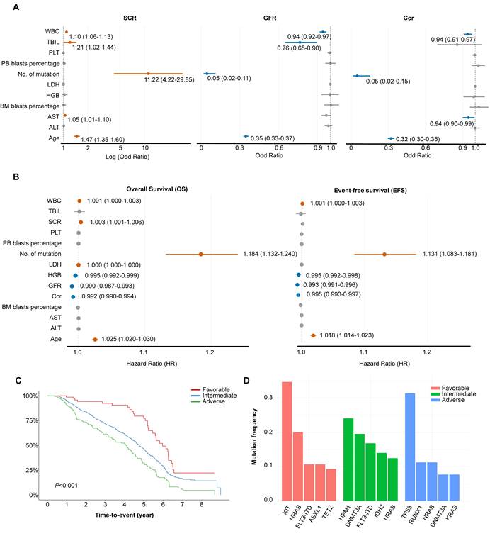
This study adhered to the principles of the Declaration of Helsinki and approved by the Expert Committee of Fujian Medical University Union Hospital in China (equivalent to an institutional review board). All participants or their guardians provided written informed consent before enrolment. AML diagnosis was established through a combination of morphology, immunology, cytogenetics and molecular biology classification following the 2008 World Health Organization[10]. This study enrolled 1,416 non-M3 AML patients admitted between 2017 and 2024 (Figure 2A). Among these, VAF data were available for 94 patients (Table S2), and protein mutation site data were available for 683 patients. We calculated Ccr using the gender-specific Cockcroft-Gault equation and GFR using the sex-adjusted CKD-EPI formula. Overall survival (OS) was measured from diagnosis to death or censoring, while event-free survival (EFS) was defined as the time to relapse, death, or censoring. Follow-up data were collected via phone, outpatient, and inpatient electronic records. Cytogenetic risk was stratified per the 2025 NCCN AML guidelines (v2.2025) (Figure 1C).
Associations between baseline characteristics and renal function/survival outcomes. (A) Logistic regression analysis showing odds ratios (ORs) for the relationship between clinical variables and renal function measures. Orange points represent statistically significant positive associations (P <0.05), blue points indicate significant negative associations, and gray points denote non-significant relationships. (B) Multi-variate cox proportional hazards model showing hazard ratios (HRs) for the relationship between clinical variables and OS/EFS. (C) Kaplan-Meier curves were stratified by prognostic risk groups based on cytogenetic classification according to the latest NCCN guidelines. (D) The top five most frequently mutated genes in each prognostic risk category according to NCCN cytogenetic stratification.
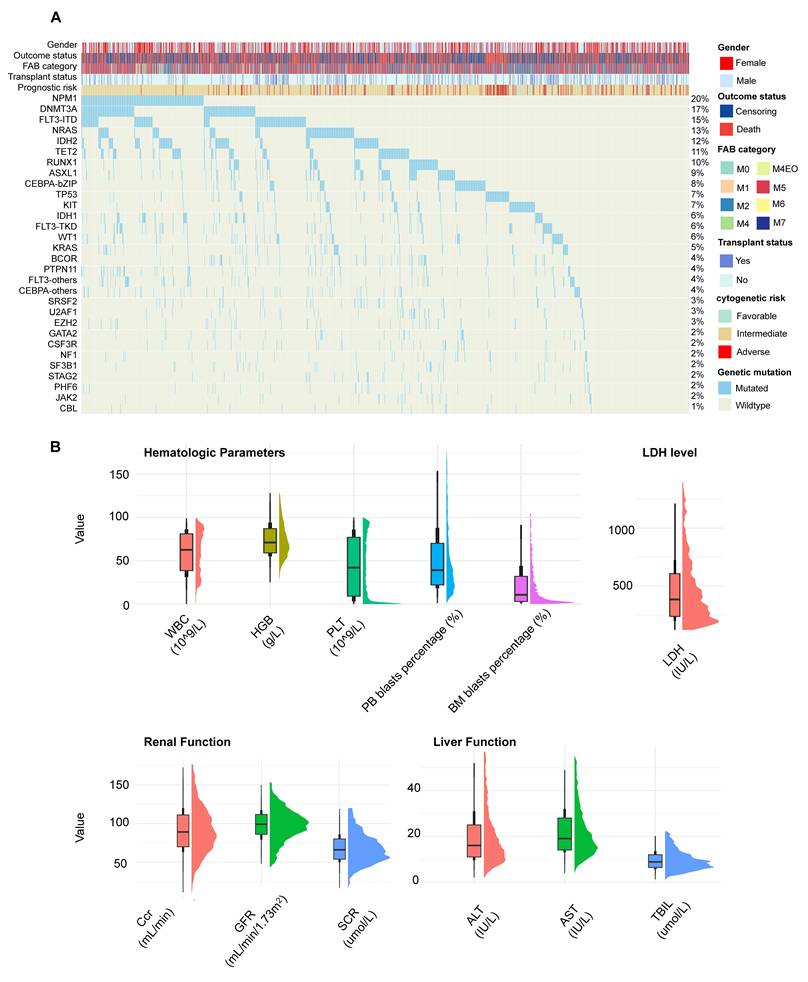
Mutational landscape and clinical summary of the patient cohort. (A) Oncoprint of genes with mutation frequencies >1% in our cohort (n = 1,416). Each column represents an individual sample, with vertical line colors indicating mutation status (mutated/unmutated). Gender, outcome, FAB subtype, transplantation status, and NCCN-defined prognostic risk are displayed above the plot. (B) Distribution and median values of clinically relevant features in the study population, stratified into four functionally distinct clusters. The Tukey boxplot method (1.5×IQR cutoff) was applied to filter outliers, which were then suppressed in subsequent plots.
Next-Generation Sequencing (NGS)
Peripheral blood of all subjects was collected in EDTA tubes. Genomic DNAs were extracted using QiaAmp Blood DNA Mini kits (Qiagen, CA, USA). The concentration and quality of genomic DNAs were assessed using a NanoDrop® ND-1000 (Thermo Fisher Scientific, MA, USA), the Qubit® 3.0 Fluorometer (Thermo Fisher Scientific, MA, USA) and 1% agarose gel electrophoresis. Paired-end DNA sequencing libraries were prepared through genomic DNA shearing using a Covaris™ (Woburn, MA, USA) sonicator, followed by peak detection, end repair, poly A-tailing, paired-end adaptor ligation, and amplification. The qualified genomic DNA sample was randomly fragmented by Covaris™ (Woburn, MA, USA) and the size of the library fragments is mainly distributed between 250bp and 300bp. End-Repair and A-tailing were then applied to facilitate ligation of the adapters, containing unique barcodes for each sample, specific to the Illumina technology for amplification and sequencing. KAPA Hyper Prep kit was used for these steps, according to the manufacturer's instructions.
Exome capture was performed using Sureselect Human All Exon V6 (Agilent Technologies, Inc., Santa Clara, CA). Each captured library was then loaded onto the NovaSeq6000 platform (Illumina, Inc., San Diego, CA). Each sample was sequenced at the mean depth of 200× to achieve high sensitivity and accuracy for mutations detection.
Target gene capture was performed using SureSelect custom designs (Agilent Technologies, Inc., Santa Clara, CA). Each captured library was loaded onto the nextseq550 platform (Illumina, Inc., San Diego, CA) at the Wuhan Kindstar Global gene Technology (Kindstar, Wuhan, China). The sequencing depth for each sample was maintained at a mean depth of 1000× for high sensitivity and accuracy in mutation detection.
Raw image files were processed using Illumina basecalling Software for base-calling with default parameters, and the sequences of each individual were generated as 150 bp pair-end reads. Pair-end reads were aligned to the human reference genome (GRCh37/HG19) using the Burrows-Wheeler Aligner (BWA, v0.7.17). Strict data quality control was performed in the entire analysis pipeline for clean data, mapping data, variant calling, etc. The Genome Analysis ToolKit (GATK, v4.1.1.0) was used for insertion/deletion realignment, quality score recalibration, and variant identification, with duplicate reads removed by the Picard-tools (v4.1.1.0). ANNOVAR (v201804) was used to annotate mutations. After sequence alignment and variant calling, synonymous variants, intronic variants far away from the exon/intron boundaries, and variants with a minor allelic frequency ≥ 1% in the 1000 Genomes Project, the dbSNP database, and the Exome Aggregation Consortium database were excluded from further analysis. NGS reads were visualized using an Integrated Genomic Viewer. Our screen covered 57 genes implicated in hematologic malignancies, grouped into seven functional clusters (Table S1). Quantitative variant allele frequency (VAF) values were available for a subset of 94 patients. All primary analyses in this study—including associations with clinical parameters, co-mutation patterns, survival models, and propensity score matching—were performed using binary mutation status, which is complete for the entire cohort. VAF data for the 94-patient subset are provided in Table S2 for readers to perform clonal abundance analyses. In addition, recurrent protein hotspot variants were summarized descriptively in patients with available protein-level variant annotation. Variant frequencies were calculated for each protein alteration, and the proportion of each hotspot among all mutations within the corresponding gene was determined.
Statistical Analysis
All statistical analyses were performed using R 4.4.1. We employed multivariate Cox proportional hazards models adjusted for age, gender, transplant status, prognostic grade[11], and FAB category to determine the prognostic impact of mutated genes. Univariate survival analyses were conducted using the Kaplan-Meier method, with results visualized via Kaplan-Meier curves and reported with log-rank p-values. To ensure balanced and comparable data, we performed propensity score matching (1:4 ratio) based on nearest-neighbor matching for age and gender prior to survival analyses. Additionally, Cohen's d values were calculated to quantify the effect sizes of different mutational patterns on clinical characteristics. To control the false-positive rate, p-values were adjusted using the Bonferroni method for analyses requiring stringent control of type I error across multiple comparisons, whereas the Benjamini-Hochberg method was applied in exploratory analyses to control the false discovery rate while maintaining statistical power. Given the limited missingness, we employed the random forest method from multiple imputation approaches to handle the missing data.
Results
Mutational Burden Defines Integrated Clinical Characteristics
Table 1 summarizes the baseline characteristics of the 1,416 enrolled patients. The cohort had a sex distribution of 42.5% female and 57.5% male. A higher mutational burden was significantly associated with older age (P < 0.001), elevated white blood cell (WBC) and platelet (PLT) counts (P < 0.001), an increased blast burden, and impaired renal function, as indicated by elevated serum creatinine (SCR), decreased GFR and Ccr (all P < 0.001). Patients with AML-M1 subtype had a significantly higher prevalence of ≥ 3 concurrent mutations compared to other subtypes (52.11% vs. 33.23%, P = 0.002). Figure S1C showed that the prognostic impact of mutational burden remained generally consistent across different clinical subgroups.
Baseline characteristic.
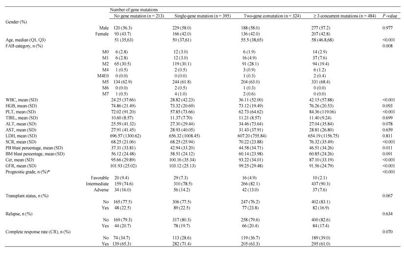
Data are presented as n (%), median (Q1, Q3), or mean (standard deviation). *The prognostic stratification was performed according to the latest NCCN 2025 guidelines, based on the patient's cytogenetic karyotypes.
Multivariate Cox analysis revealed that advanced age, higher mutational burden, and elevated WBC counts were independently associated with impaired renal function (Figure 1A). And these factors together with damaged renal function independently predict poor prognosis in AML patients (Figure 1B).
Mutation-Specific Organ Toxicity and Prognostic Refinement
The mutation landscape shows genes mutated in > 1% of the cohort (Figure 2A). The most frequently mutated gene in AML was NPM1 (n = 285, 20%), followed by DNMT3A (n = 243, 17%) and FLT3-ITD (n = 215, 15%) (Figure 2A). To further characterize recurrent mutations within specific genes, we examined the distribution of protein variants in the subset of 683 patients with available protein-level annotation. Consistent with previous findings[12-21], the identified mutations were predominantly located at well-established hotspot residues of AML-associated genes, including NPM1 p.W288Cfs*12, IDH2 p.R140Q, DNMT3A p.R882H, NRAS p.G12D, FLT3 p.D835Y, and KIT p.D816V. The distribution of these recurrent hotspot variants is summarized in Table S3. Consistent with previous reports[22-25], mutations in DNMT3A, TET2, TP53, SRSF2, RUNX1, ASXL1, and NPM1 were more prevalent in older patients, whereas CEBPA-bZIP and KIT mutations predominated in younger patients (Figure S2A). Consistent with previous studies, RUNX1 mutations showed male predominance[26, 27], contrasting with the female predominance of FLT3-ITD [28]. TP53 (P = 0.025) and CEBPA-bZIP (P = 0.022) mutations were significantly more prevalent in males, while IDH1 was more frequent in females (P = 0.014). Nonetheless, NPM1 was the most frequently mutated gene in both genders (Figure S1A). In our cohort, karyotypes separated patients into three groups with significantly different prognosis (Figure 1C). In line with prior reports[29], KIT mutations were most frequent in patients with favorable-risk cytogenetics (34.67%). The intermediate-risk group was characterized by predominant NPM1 (19.96%) and DNMT3A (15.67%) mutations, whereas the adverse-risk group showed a high prevalence of TP53 (31.36%) and RUNX1 (11.24%) mutations (Figure 1D). Analysis of AML subtypes revealed significantly higher frequencies of NPM1 (35.78%) and IDH2 (26.61%) mutations in the M0/M1 subtype compared to others (P < 0.001 for both; Figure S1B).
Beyond the mutational frequencies across demographic and clinical subgroups, we next investigated the correlations between specific mutations and key laboratory parameters. TP53, BCOR, IDH2, and U2AF1 mutations correlated with low WBC counts, while FLT3-ITD, NPM1, NRAS, DNMT3A, and FLT3-others were associated with high WBC counts[23, 30, 31](Figure S2B). DNMT3A mutations additionally correlated with elevated PLT counts (Figure S2C). Consistent with previous reports[23, 32-34], TP53 and ASXL1 mutations showed higher prevalence in patients with low bone marrow (BM) and peripheral blood (PB) blast percentage (Figure S4B-C). FLT3-ITD, NPM1, DNMT3A, IDH1, and IDH2 mutations dominated high BM blast groups, while FLT3-ITD, NPM1, and CEBPA-bZIP mutations characterized high PB blasts percentage groups[23, 35-38]. Contrary to prior reports indicating no significant correlation between RUNX1 mutation and BM blast percentage[39], patients harboring RUNX1 mutations exhibited reduced BM and PB blasts compared to wild-type cases (Figure S4B-C).
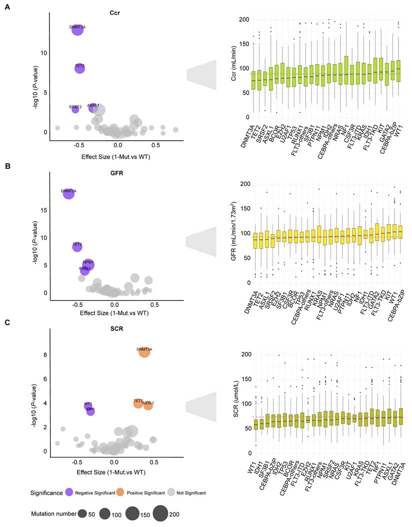
Given limited research on mutations linked to hepatic and renal impairment in AML, we evaluated specific mutation effects. DNMT3A, TET2, and ASXL1 mutations correlated with renal impairment (reduced Ccr and GFR, elevated SCR; Figure 3A-C). Renal impairment was defined as elevated SCR or a significant reduction in GFR (P < 0.05). Multivariate regression analysis confirmed DNMT3A mutations as an independent factor for all renal impairment indicators (GFR, Ccr and SCR), while TET2 mutations independently affected Ccr and ASXL1 mutations affected GFR and SCR (Figure S3B). Although the effects of most mutations on liver function are generally marginal, FLT3-ITD or NPM1 mutations correlated with elevated aspartate aminotransferase (AST) (Figure S5A), and CEBPA-bZIP with increased total bilirubin (TBIL) (Figure S5C). Similarly, IDH2, RUNX1, and BCOR mutations correlated with low lactate dehydrogenase (LDH) while FLT3-ITD and KIT mutations associated with high LDH (Figure S3A).
Association between individual genotypes and renal functions of AML. Statistical correlation (left) and distribution (right) between renal function indicators (SCR, GFR and Ccr) and individual genotypes for the patients in our cohort. Left panels: Orange points indicate statistically significant (Bonferroni FDR <0.05) positive effect sizes while purple points indicate statistically negative effect sizes. Effect sizes (Cohen's d) were calculated between mutated and wild-type patients. P-values were calculated using a two-sided Wilcoxon rank sum test. Right panels: Red dashed lines denote the mean value for each feature across all patients. For every distribution, boxplots display the first and third quartile boundaries, with a central line marking the median in different gene-mutated patients.
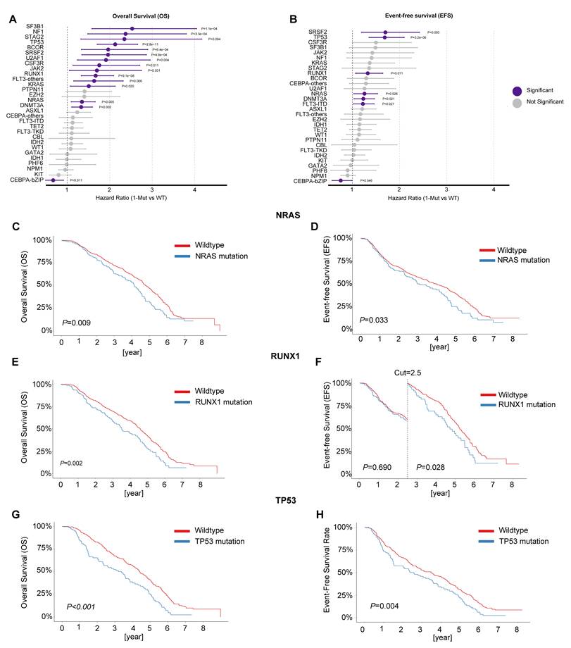
Having established the clinical and laboratory correlates of specific mutations, we next evaluated their prognostic significance through multivariable Cox proportional hazards regression models. Consistent with prior studies[40-46], RUNX1, DNMT3A, SRSF2, NRAS, and TP53 mutations were associated with significantly adverse OS and EFS (Figure 4A-H). CSF3R mutations predicted adverse OS across all AML subtypes, extending its previously limited prognostic association with CEBPA-mutant AML[47] (Figure S6E). Additionally, we validated the favorable OS (Figure S6J) and EFS (Figure S7C) in AML patients with CEBPA-bZIP mutations (HR = 0.67, 95%; CI: 0.49-0.91) (Figure 4A-B). However, NF1, CSF3R, JAK2, KRAS, STAG2, U2AF1, BCOR, and FLT3-others only impacted OS without affecting EFS (Figure 4A-B).
Association between individual genotypes and survival outcomes of AML. (A) Forestplot depicting multivariate Cox proportional hazards analysis of overall survival for genes with mutation frequencies >1% in the study cohort, adjusted for age, gender, and transplantation status. Genes demonstrating statistical significance (P <0.05) are highlighted in purple. (B) Forestplot depicting multivariate Cox proportional hazards analysis of event-free survival (events defined as all-cause death or relapse) for genes with >1% mutation frequency in the study population, adjusted for age, gender, and transplantation status. Genes reaching statistical significance (P <0.05) are shown in purple. (C-H) Kaplan-Meier survival curves for the mutation of NRAS, RUNX1 and TP53. Landmark analysis was applied where curves crossed.
Co-mutation Networks Drive Adverse Outcomes and Organ Toxicity
We comprehensively characterized the co-mutation landscape of recurrently altered genes (mutation frequency > 1%) in the entire cohort. Beyond previously reported associations (e.g., NPM1/PTPN11, and SRSF2/STAG2)[23], we identified several novel co-occurrences (e.g., KRAS and PTPN11; OR = 3.19; 95% CI, 2.97-3.42; adjusted P < 0.001) or mutually exclusive (e.g., CEBPA-bZIP and NPM1; OR = 0.03; 95% CI, 0.01-0.17; adjusted P <0.001) gene pairs (Figure 5A; Figure S8A).
Distinct patterns of mutation co-occurrence associate with overall survival and clinical characteristics. (A) Co-occurrence and mutual exclusivity of the most frequent mutations present in de novo AML was performed using a two-sided Fisher's Exact test. Point size and color are based on the magnitude and directionality of the odds ratio based on co-occurrence (red) or mutual exclusivity (blue). Asterisks representing the FDR-corrected significance. (B) Summary of pairwise mutations and their association with prognosis based on Cox proportional-hazards regression modeling adjusted for age, gender, and transplantation status. Significant genotypes (Bonferroni FDR <0.05) are colored according to the log-transformed hazard ratio compared to wild-type patients, with purple representing worse prognosis (HR ≥ 1). Points are sized based on the number of patients with co-occurring mutations for each genotype. (C-H) Impact of genetic pair mutations on patient clinical characteristics. Orange points indicate statistically significant (Bonferroni FDR <0.05) positive effect sizes while purple points indicate statistically negative effect sizes. Effect sizes (Cohen's d) were calculated between mutated and wild-type patients. P-values were calculated using a two-sided Wilcoxon rank sum test.
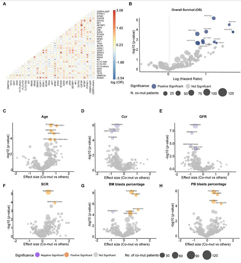
Analysis of the association between co-mutation patterns and age revealed that several co-mutations SRSF2/ASXL1, TET2/NPM1, and TET2/ASXL1 were enriched in older patients (Figure 5C). Notably, patients with DNMT3A/NPM1 or DNMT3A/FLT3-others co-mutations exhibited significantly elevated SCR, decreased GFR, and reduced Ccr (Figure 5D-F), although these combinations did not confer worsened renal function compared to DNMT3A mutations alone. We also assessed the relationship between co-mutation patterns and liver function markers (alanine aminotransferase (ALT), AST, TBIL) and LDH. No co-mutation combinations were significantly associated with liver function parameters (Figure S8C-E).
Consistent with previous study[23], we validated that NPM1/DNMT3A, NPM1/FLT3-ITD, and DNMT3A/FLT3-ITD co-mutations were associated with elevated WBC counts, which may contribute to their poor prognosis. Furthermore, IDH2/BCOR co-mutations were associated with significantly lower WBC counts (Figure S8F). Co-mutations of NPM1 with IDH1, IDH2, or FLT3-ITD were associated with increased blast proportions (Figure 5G-H). Given that single mutations in these genes were all associated with increased leukemic cell burden, we evaluated whether co-mutations exhibited additive effects relative to single mutations. The NPM1/IDH1 co-mutation significantly increased BM and PB blast percentages compared to NPM1 single mutation (β = 16.797 for BM blasts percent, P < 0.001; β = 22.101 for PB blast percentages, P < 0.001) or IDH1 single mutation (β = 22.097 for BM blasts percent, P < 0.001; β = 35.032 for PB blasts percent, P < 0.001). Similarly, NPM1/FLT3-ITD co-mutations showed significantly increased blasts compared to NPM1 single mutation (β = 8.790 for BM blasts percent, P = 0.004; β = 10.368 for PB blasts percent, P = 0.014), though not compared to FLT3-ITD single mutation. Additionally, NPM1/IDH2 co-mutations significantly increased blasts compared to IDH2 single mutation (β = 9.751 for BM blasts percent, P = 0.007; β = 26.648 for PB blasts percent, P < 0.001), but not compared to NPM1 single mutation.
All significant co-occurring mutations were associated with adverse prognosis (Figure 5B). Consistent with prior studies[48, 49], ASXL1 co-mutations with RUNX1 (HR = 2.23, adjust P < 0.001) or SRSF2 (HR = 3.27, adjusted P < 0.001) were associated with significantly worse outcomes (Figure 5B).
Specific Triple Mutations Predict Renal Impairment and Poorer Survival
We identified 30 distinct triple-mutation genotypes (≥ 7 patients each; Figure 6A). Four combinations exhibited significantly higher co-occurrence frequencies than random expectation: IDH2: FLT3-ITD: NPM1, IDH2: SRSF2: ASXL1, RUNX1: TET2: NRAS and DNMT3A: IDH2: FLT3-ITD (Figure 6A). We then analyzed the impact of these four triple-mutation combinations on clinical characteristics. We detected the DNMT3A: IDH2: FLT3-ITD mutation combination demonstrated significantly reduced WBC counts (Figure 6L). Notably, patients harboring the RUNX1: TET2: NRAS triple mutation exhibited significantly reduced GFR and Ccr (Figure 6I). However, no significant associations were observed between these triple-mutation genotypes and patient age.
Mutational landscape of triple mutations and their associations with survival and clinical characteristics. (A) Frequency distribution of the number of de novo patients with the most frequent 3-way mutation combinations. Bars are colored based on the association with a significant survival correlation (P ≤ 0.05) compared to patients with only two genes mutated: red = a significant survival association, gray = no significant association. (B) Forestplot depicting survival analysis between triple-mutated and double-mutated genotypes. Points represent the hazard ratios calculated between triple vs. double-mutated patients using a Cox proportional-hazards model, adjusted for age, gender, and transplantation status. Significant genotypes are colored: purple points represent genotypes where all three mutations correlated with worse survival. Points are sized relative to the number of patients with all three mutations and bars represent the 95% confidence intervals of the hazard ratios. (C-E) Impact of IDH2: FLT3-ITD: NPM1 co-mutation on survival and clinical phenotypes in AML. The point of forestplot is colored based on the level of significance, the purple point indicates genes meeting Bonferroni FDR <0.1, the green point represents those with Bonferroni FDR <0.3, while the gray shows no statistical significance; bars represent the 95% confidence intervals of the effect sizes. Overall survival and event-free survival were depicted using Kaplan-Meier survival curves, with landmark analysis applied at the time point where the curves crossed. (F-H) Impact of IDH2: SRSF2: ASXL1 co-mutation on survival and clinical phenotypes in AML. (I-K) Impact of RUNX1: TET2: NRAS co-mutation on survival and clinical phenotypes in AML. (L-N) Impact of DNMT3A: IDH2: FLT3-ITD co-mutation on survival and clinical phenotypes in AML. Overall survival and event-free survival were depicted using Kaplan-Meier survival curves, with landmark analysis applied at the time point where the curves crossed.
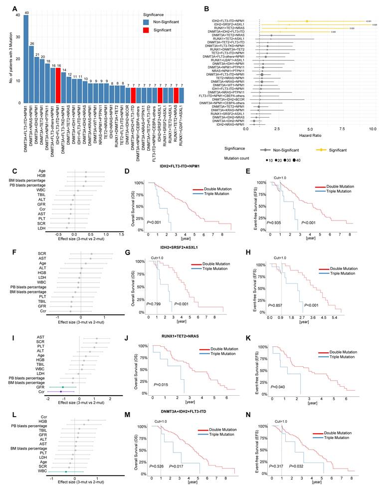
In the study by Dunlap et al[50], compared to NPM1-mutated/FLT3-ITD-negative cases, patients with concurrent DNMT3A and IDH1/2 mutations in addition to NPM1 (triple-positive) exhibited significantly shorter OS. This indicates that these specific co-occurring mutations abrogate the favorable prognosis conferred by isolated NPM1 mutation. We augmented our analysis to investigate the prognostic association of triple-mutated genotypes compared to pairwise mutated genotypes. Beyond established adverse effects of NPM1/FLT3-ITD co-mutations (per ELN 2022/NCCN 2025 guidelines), the IDH2: FLT3-ITD: NPM1 triple mutation markedly worsened survival (HR = 4.68, 95% CI = (2.39-9.15), adjusted P < 0.001). Consistent with previous report[23], DNMT3A: IDH2: FLT3-ITD (HR = 2.61, 95% CI = (1.05-6.49) adjusted P = 0.040) were associated with worse outcomes (Figure 6B). In addition, we identified another two novel high-risk triple-mutations: IDH2: SRSF2: ASXL1 (HR = 3.31, 95% CI = (1.13-9.68), adjusted P = 0.028), and RUNX1: TET2: NRAS (HR = 3.08, 95% CI = (1.20-7.92) adjusted P = 0.020) (Figure 6B). All significantly reduced OS and EFS (Figure 6D-E, G-H, J-K, M-N). The observed renal impairment in RUNX1: TET2: NRAS triple mutation may contribute to its poor prognosis. The DNMT3A: IDH2: FLT3-ITD mutation combination was associated with leukopenia. It's unknown whether and how reduced WBC counts would impact survival outcome.
Discussion
Our study demonstrates that high-frequency co-mutation modules (dual/triple-hit) are associated with AML progression, linking mutational accumulation to clonal evolution in 1,416 patients. This moves beyond single-driver genetics. The accumulation of age-related mutations underscores clonal hematopoiesis of indeterminate potential (CHIP) as a leukemogenic precursor[51]. Early recognition of CHIP mutations may enable proactive monitoring for subsequent leukemia driver mutations. Our study suggested the high-risk co-mutations (e.g., ASXL1-SRSF2, TET2-BCOR) drive leukemogenesis through genetic cooperation and predict poor prognosis. These findings illuminate critical interception points for monitoring CHIP-related genetic evolution and preventing progression to overt leukemia, supporting tailored surveillance strategies.
Our analysis identified four high-risk triple-mutation combinations. Notably, the RUNX1: TET2: NRAS combination was associated with renal dysfunction, which may contribute to its poor outcomes. Despite the conventional association of low WBC with favorable outcomes, the DNMT3A: IDH2: FLT3-ITD triple-mutant cohort paradoxically exhibited both reduced leukocyte counts and inferior survival. This apparent contradiction may reflect a stealth aggression phenotype. IDH2-derived 2-HG suppresses mitochondrial oxidative phosphorylation[52, 53], diminishing blast proliferation without impairing survival—enabling chemoresistance through stress adaptation. DNMT3A mutations drive aberrant extramedullary hematopoiesis and create a permissive epigenetic state, while FLT3-ITD signaling amplifies proliferative signals[54, 55], facilitating bone marrow niche colonization and extramedullary invasion despite low peripheral blasts.
Beyond the clinical associations described above, the co-occurrence frequencies of these four triple mutation combinations significantly exceeded random expectation, suggesting non-random selective pressures. We propose the following synergistic mechanisms.
For the IDH2: FLT3-ITD: NPM1 combination, IDH2-derived 2-HG may impair dendritic cell differentiation and reduce MHC class II expression, creating an immunosuppressive niche[56]; we hypothesize that this protects NPM1-mutant clones, which otherwise elicit neoepitope-specific T-cell responses, from immune elimination[57]. Meanwhile, FLT3-ITD drives constitutive proliferation and, together with NPM1 mutation, remodels chromatin accessibility to enforce a differentiation-blocked state[4, 58, 59]. Thus, immune evasion, proposed protection of an immunogenic clone, and proliferative/differentiation signals represent three complementary hits that no single or double mutation can fully provide.
Turning to IDH2: SRSF2: ASXL1, preclinical evidence shows that Asxl1 truncation plus Srsf2 mutation drives leukemogenesis via immune reprogramming[60]; adding IDH2 may further disable antigen presentation. The three mutations converge on distinct layers of gene regulation: IDH2 causes DNA hypermethylation and altered histone methylation[61-63]; SRSF2 disrupts splicing of key hematopoietic regulators[21]; ASXL1 impairs H3K27 trimethylation[64]. Their co-occurrence likely reflects a need to simultaneously disrupt multiple epigenetic barriers—a task unachievable by any single mutation.
In the RUNX1: TET2: NRAS combination, RUNX1 is a master regulator of myeloid differentiation[65]. RUNX1 recruits TET2 to mediate site-specific DNA demethylation[66]. TET2 loss compromises DNA methylation fidelity and has been linked to hematopoietic dysfunction[67-69]. Their concurrent loss may synergistically disrupt epigenetic regulation, while NRAS activation supplies a constitutive proliferative driver. This triad thus may evolve through acquisition of hits that abrogate differentiation (RUNX1), epigenetic fidelity (TET2), and accelerated proliferation (NRAS).
For DNMT3A: IDH2: FLT3-ITD, the co-occurrence may follow a combinatorial evolutionary trajectory: DNMT3A mutation as an epigenetic primer that alters the methylation landscape[54]; IDH2 mutation as a metabolic and immune modulator that suppresses oxidative phosphorylation and T-cell function[52, 56]; and FLT3-ITD as a transforming hit that amplifies proliferative signals[59]. This co-occurrence pattern, rather than random co-occurrence, may enable immune evasion, metabolic adaptation, and niche colonization despite low peripheral blast counts.
Collectively, these four combinations illustrate distinct but convergent mechanisms- functional complementation, clonal evolution, and multi-layered epigenetic/immune disruption—that may drive their non-random co-occurrence and adverse prognosis.
A higher mutational burden was associated with impaired renal function indicated by GFR and CCr. Impaired renal function was significantly associated with both single mutations (DNMT3A, TET2, or ASXL1) and, with the DNMT3A and NPM1 co-mutation, suggesting a potential synergistic effect on renal pathology.
We propose a distinct mechanism wherein DNMT3A dysfunction induces PTEN promoter hypermethylation, suppressing PTEN expression and activating the PI3K/AKT pathway[70, 71]. This epigenetic dysregulation accelerates renal tubular EMT and TGF-β1-driven fibrotic signaling, exacerbating renal damage[72].
While DNMT3A mutation was independently associated with renal impairment in our multivariable model, we acknowledge that residual confounding by age, sex, and cytogenetic risk may exist. Given the retrospective observational design, this association does not imply causation, and prospective studies are needed to validate this finding.
Collectively, these findings may underscore the need to integrate mutational profiling with renal function monitoring. Prospective validation is warranted to operationalize this precision nephroprotection paradigm.
This study establishes a foundational framework elucidating the genomic-clinical interplay in AML[73, 74]. Proactive surveillance of patients with clonal hematopoiesis (CH), coupled with timely intervention of high-risk co-mutations, may facilitate early detection of leukemic transformation[75]. However, this study also has several limitations. Reliable VAF data were available for only 94 of 1,416 patients. Consequently, we were unable to assess the impact of variant allele frequency on prognosis or clonal architecture in the majority of the cohort. While overall sampling was robust, analyses of rare co-mutations or phenotypes were underpowered. Furthermore, the exclusive Chinese cohort enhances homogeneity but may limit global applicability due to known ethnic disparities in AML.
Future studies should develop CH risk models incorporating high-risk co-mutations and renal impairment biomarkers, while optimizing therapies via renal-adapted, mutation-informed drug combinations that avoid nephrotoxicity. Real-time organ surveillance using biomarker panels and toxicity predictors must be implemented. These strategies require validation through multi-center consortia across diverse cohorts to advance precision interception in AML.
Conclusion
Co-mutation patterns are pivotal in shaping AML phenotypes and outcomes. Our findings highlight the value of integrating genomic and biochemical data, particularly renal function metrics, to improve risk assessment. These findings also support the development of tailored management strategies that address both the leukemia itself and its associated organ toxicities.
Abbreviations
ALT: alanine aminotransferase; AST: aspartate aminotransferase; BM: bone marrow; Ccr: creatinine clearance rate; GFR: glomerular filtration rate; HGB: hemoglobin; LDH: lactate dehydrogenase; NGS: Next-generation sequencing; PB: peripheral blood; PLT: platelet; SCR: serum creatinine; TBIL: total bilirubin; VAF: variant allele frequency; WBC: white blood cell.
Supplementary Material
Supplementary figures and tables.

Acknowledgements
We thank all the AML patients for their participation in this study.
Funding
This work was supported by the National Natural Science Foundation of China (82170148), the Excellent Young Scientists Project of Fujian Provincial Natural Science Foundation (2025J09040), Joint Funds for the Innovation of Science and Technology of Fujian province (2021Y9052), Excellent Young Scholars Cultivation project of Fujian Medical University Union Hospital (2022XH023) , as well as National Key Clinical Specialty Discipline Construction Program (2021-76) and Fujian Provincial Clinical Research Center for Hematological Malignancies (2020Y2006). And we thank all the AML patients for their participation.
Ethics statement
This study was conducted in accordance with the ethical standards of the Helsinki Declaration. The study protocol was approved by the ethics committee of Fujian Medical University Union Hospital. All patients have given written informed consent.
Consent for publication
Informed consent was obtained from all individual participants included in the study.
Data availability
The datasets generated during and/or analyzed during the current study are not publicly available due to ethical restrictions but are available from the corresponding author, Lili Pan (theresalilipan@fjmu.edu.cn), upon reasonable request. The computational code used in this study can also be obtained from the corresponding author upon reasonable request.
Author Contributions
ZX was responsible for statistical analysis of clinical data and for writing and revising the manuscript. YZ was responsible for collating clinical data and for writing and revising the manuscript. YZ (a different author with the same initials) collected the clinical data. XL contributed to the study design by providing critical suggestions. PL conceived and designed the study, supervised the research, revised the manuscript, and acquired the funding. SW supervised the project and provided guidance.
Competing Interests
The authors have declared that no competing interest exists.
References
1. Newell LF, Cook RJ. Advances in acute myeloid leukemia. BMJ. 2021;375:n2026
2. Khoury JD, Solary E, Abla O, Akkari Y, Alaggio R, Apperley JF. et al. The 5th edition of the World Health Organization Classification of Haematolymphoid Tumours: Myeloid and Histiocytic/Dendritic Neoplasms. Leukemia. 2022;36:1703-19
3. Zhang S, Huang F, Wang Y, Long Y, Li Y, Kang Y. et al. NAT10-mediated mRNA N4-acetylcytidine reprograms serine metabolism to drive leukaemogenesis and stemness in acute myeloid leukaemia. Nature Cell Biology. 2024;26:2168-82
4. Yun H, Narayan N, Vohra S, Giotopoulos G, Mupo A, Madrigal P. et al. Mutational synergy during leukemia induction remodels chromatin accessibility, histone modifications and three-dimensional DNA topology to alter gene expression. Nat Genet. 2021;53:1443-55
5. Lachowiez CA, Long N, Saultz J, Gandhi A, Newell LF, Hayes-Lattin B. et al. Comparison and validation of the 2022 European LeukemiaNet guidelines in acute myeloid leukemia. Blood Adv. 2023;7:1899-909
6. Dohner H, Wei AH, Appelbaum FR, Craddock C, DiNardo CD, Dombret H. et al. Diagnosis and management of AML in adults: 2022 recommendations from an international expert panel on behalf of the ELN. Blood. 2022;140:1345-77
7. Arber DA, Orazi A, Hasserjian RP, Borowitz MJ, Calvo KR, Kvasnicka HM. et al. International Consensus Classification of Myeloid Neoplasms and Acute Leukemias: integrating morphologic, clinical, and genomic data. Blood. 2022;140:1200-28
8. Tazi Y, Arango-Ossa JE, Zhou Y, Bernard E, Thomas I, Gilkes A. et al. Unified classification and risk-stratification in Acute Myeloid Leukemia. Nat Commun. 2022;13:4622
9. Jiang X, Mak PY, Mu H, Tao W, Mak DH, Kornblau S. et al. Disruption of Wnt/beta-Catenin Exerts Antileukemia Activity and Synergizes with FLT3 Inhibition in FLT3-Mutant Acute Myeloid Leukemia. Clin Cancer Res. 2018;24:2417-29
10. Vardiman JW, Thiele J, Arber DA, Brunning RD, Borowitz MJ, Porwit A. et al. The 2008 revision of the World Health Organization (WHO) classification of myeloid neoplasms and acute leukemia: rationale and important changes. Blood. 2009;114:937-51
11. Wang P, Cai Q, Peng X, Dai Z, Liu J, Li W. et al. Increased co-expression of CTLA4/LAG3 predicted adverse clinical outcomes in patients with T-cell malignancies. Cell Investigation. 2025 1
12. Bailey E, Li L, Duffield AS, Ma HS, Huso DL, Small D. FLT3/D835Y mutation knock-in mice display less aggressive disease compared with FLT3/internal tandem duplication (ITD) mice. Proceedings of the National Academy of Sciences. 2013;110:21113-8
13. Giannakopoulou E, Lehander M, Virding Culleton S, Yang W, Li Y, Karpanen T. et al. A T cell receptor targeting a recurrent driver mutation in FLT3 mediates elimination of primary human acute myeloid leukemia in vivo. Nature Cancer. 2023;4:1474-90
14. Takiar V, Ip CKM, Gao M, Mills GB, Cheung LWT. Neomorphic mutations create therapeutic challenges in cancer. Oncogene. 2016;36:1607-18
15. Ward PS, Patel J, Wise DR, Abdel-Wahab O, Bennett BD, Coller HA. et al. The Common Feature of Leukemia-Associated IDH1 and IDH2 Mutations Is a Neomorphic Enzyme Activity Converting α-Ketoglutarate to 2-Hydroxyglutarate. Cancer Cell. 2010;17:225-34
16. Marcucci G, Metzeler KH, Schwind S, Becker H, Maharry K, Mrózek K. et al. Age-Related Prognostic Impact of Different Types of DNMT3A Mutations in Adults with Primary Cytogenetically Normal Acute Myeloid Leukemia. Journal of Clinical Oncology. 2012;30:742-50
17. Ley TJ, Ding L, Walter MJ, McLellan MD, Lamprecht T, Larson DE. et al. DNMT3AMutations in Acute Myeloid Leukemia. New England Journal of Medicine. 2010;363:2424-33
18. Yuan R, Xie Y, Miron PM, Higgins AW, Hutchinson L, Cerny J. et al. Clinico-genomic characterization of RAS-mutant acute myeloid leukemia. Annals of Hematology. 2026 105
19. Zhao L, Melenhorst JJ, Alemu L, Kirby M, Anderson S, Kench M. et al. KIT with D816 mutations cooperates with CBFB-MYH11 for leukemogenesis in mice. Blood. 2012;119:1511-21
20. Cairoli R. Prognostic impact of c-KIT mutations in core binding factor leukemias: an Italian retrospective study. Blood. 2006;107:3463-8
21. Yu H, Hong J, Shin D-Y, Lee C-H. The role of ASXL1, SRSF2, and EZH2 mutations in chromatin dysregulation of myelodysplastic neoplasia and acute myeloid leukemia. Leukemia. 2025;39:2329-39
22. Dou X, Dan C, Zhang D, Zhou H, He R, Zhou G. et al. Genomic mutation patterns and prognostic value in de novo and secondary acute myeloid leukemia: A multicenter study from China. Int J Cancer. 2024;155:2253-64
23. Benard BA, Leak LB, Azizi A, Thomas D, Gentles AJ, Majeti R. Clonal architecture predicts clinical outcomes and drug sensitivity in acute myeloid leukemia. Nat Commun. 2021;12:7244
24. Straube J, Ling VY, Hill GR, Lane SW. The impact of age, NPM1mut, and FLT3ITD allelic ratio in patients with acute myeloid leukemia. Blood. 2018;131:1148-53
25. Georgi JA, Stasik S, Kramer M, Meggendorfer M, Röllig C, Haferlach T. et al. Prognostic impact of CEBPA mutational subgroups in adult AML. Leukemia. 2024;38:281-90
26. Gaidzik VI, Teleanu V, Papaemmanuil E, Weber D, Paschka P, Hahn J. et al. RUNX1 mutations in acute myeloid leukemia are associated with distinct clinico-pathologic and genetic features. Leukemia. 2016;30:2160-8
27. Hellesøy M, Engen C, Grob T, Löwenberg B, Valk PJM, Gjertsen BT. Sex disparity in acute myeloid leukaemia with FLT3 internal tandem duplication mutations: implications for prognosis. Molecular Oncology. 2021;15:2285-99
28. Höglund M, Lazarevic VL, Ölander E, Lorenz F, Cammenga J, Antunovic P. et al. The prognostic impact of FLT3-ITD and NPM1 mutation in adult AML is age-dependent in the population-based setting. Blood Advances. 2020;4:1094-101
29. Li L, Ruan J, Zhang N, Dai J, Xu X, Tian X. et al. Identification of prognostic and driver gene mutations in acute myeloid leukemia by a bioinformatics analysis. Translational Cancer Research. 2023;12:1552-64
30. Zhou Y, Zhang H, Zheng X, Zhang R, Chen X, Ma Q. et al. Impact of BCOR/BCORL1 mutation on outcomes of allogeneic hematopoietic stem cell transplantation in acute myeloid leukemia patients. Ann Hematol. 2025;104:2631-42
31. Gao Y, Jia M, Mao Y, Cai H, Jiang X, Cao X. et al. Distinct Mutation Landscapes Between Acute Myeloid Leukemia With Myelodysplasia-Related Changes and De Novo Acute Myeloid Leukemia. Am J Clin Pathol. 2022;157:691-700
32. Yang L, Wei X, Gong Y. Prognosis and risk factors for ASXL1 mutations in patients with newly diagnosed acute myeloid leukemia and myelodysplastic syndrome. Cancer Med. 2024;13:e6871
33. Park S, Kim S-A, Kim S, Kim SY, Lee JY, Byun JM. et al. Distinct immunophenotypic and clinical features of TP53-mutated acute myeloid leukemia: high CD34/CD41 expression and lower leukocyte counts. Annals of Hematology. 2026 105
34. Welch JS. Patterns of mutations in TP53 mutated AML. Best Practice & Research Clinical Haematology. 2018;31:379-83
35. Notopuro P, Nugraha J, Utomo B, Notopuro H. The Association of FLT3-ITD Gene Mutation with Bone Marrow Blast Cell Count, CD34, Cyclin D1, Bcl-xL and hENT1 Expression in Acute Myeloid Leukemia Patients. Iranian Journal of Pathology. 2020;15:306-12
36. Yang X, Shi J, Zhang X, Zhang G, Zhang J, Yang S. et al. Biological and clinical influences of NPM1 in acute myeloid leukemia patients with DNMT3A mutations. Cancer Manag Res. 2018;10:2489-97
37. Zarnegar-Lumley S, Alonzo TA, Gerbing RB, Othus M, Sun Z, Ries RE. et al. Characteristics and prognostic impact of IDH mutations in AML: a COG, SWOG, and ECOG analysis. Blood Adv. 2023;7:5941-53
38. Tien F-M, Yao C-Y, Tsai XC-H, Lo M-Y, Chen C-Y, Lee W-H. et al. Dysregulated immune and metabolic pathways are associated with poor survival in adult acute myeloid leukemia with CEBPA bZIP in-frame mutations. Blood Cancer Journal. 2024 14
39. Khan M, Cortes J, Kadia T, Naqvi K, Brandt M, Pierce S. et al. Clinical Outcomes and Co-Occurring Mutations in Patients with RUNX1-Mutated Acute Myeloid Leukemia. Int J Mol Sci. 2017 18
40. Rungjirajittranon T, Siriwannangkul T, Kungwankiattichai S, Leelakanok N, Rotchanapanya W, Vittayawacharin P. et al. Clinical Outcomes of Acute Myeloid Leukemia Patients Harboring the RUNX1 Mutation: Is It Still an Unfavorable Prognosis? A Cohort Study and Meta-Analysis. Cancers (Basel). 2022 14
41. Park DJ, Kwon A, Cho BS, Kim HJ, Hwang KA, Kim M. et al. Characteristics of DNMT3A mutations in acute myeloid leukemia. Blood Res. 2020;55:17-26
42. van der Werf I, Wojtuszkiewicz A, Meggendorfer M, Hutter S, Baer C, Heymans M. et al. Splicing factor gene mutations in acute myeloid leukemia offer additive value if incorporated in current risk classification. Blood Adv. 2021;5:3254-65
43. Alawieh D, Cysique-Foinlan L, Willekens C, Renneville A. RAS mutations in myeloid malignancies: revisiting old questions with novel insights and therapeutic perspectives. Blood Cancer J. 2024;14:72
44. Ni SC, Yao CY, Tsai XC, Lo MY, Chen CY, Lee WH. et al. Genomic and transcriptomic determinants of clinical outcomes in patients with AML and DNMT3A mutations. Blood Cancer J. 2025;15:97
45. Qin G, Han X. The Prognostic Value of TP53 Mutations in Adult Acute Myeloid Leukemia: A Meta-Analysis. Transfusion Medicine and Hemotherapy. 2023;50:234-44
46. Jin H, Zhu Y, Hong M, Wu Y, Qiu H, Wang R. et al. Co-occurrence of KIT and NRAS mutations defines an adverse prognostic core-binding factor acute myeloid leukemia. Leukemia & Lymphoma. 2021;62:2428-37
47. Meshinchi S, Maxson JE, Smith JL, Hylkema T, Ries RE, Gerbing RB. et al. Prognostic impact of CSF3R mutations in favorable risk childhood acute myeloid leukemia. Blood. 2020;135:1603-6
48. Sui P, Ge G, Chen S, Bai J, Rubalcava IP, Yang H. et al. SRSF2 mutation cooperates with ASXL1 truncated alteration to accelerate leukemogenesis. Leukemia. 2023;38:408-11
49. Paschka P, Schlenk RF, Gaidzik VI, Herzig JK, Aulitzky T, Bullinger L. et al. ASXL1 mutations in younger adult patients with acute myeloid leukemia: a study by the German-Austrian Acute Myeloid Leukemia Study Group. Haematologica. 2015;100:324-30
50. Dunlap JB, Leonard J, Rosenberg M, Cook R, Press R, Fan G. et al. The combination of NPM1, DNMT3A, and IDH1/2 mutations leads to inferior overall survival in AML. American Journal of Hematology. 2019;94:913-20
51. Jahn E, Saadati M, Fenaux P, Gobbi M, Roboz GJ, Bullinger L. et al. Clinical impact of the genomic landscape and leukemogenic trajectories in non-intensively treated elderly acute myeloid leukemia patients. Leukemia. 2023;37:2187-96
52. Fu X, Chin Randall M, Vergnes L, Hwang H, Deng G, Xing Y. et al. 2-Hydroxyglutarate Inhibits ATP Synthase and mTOR Signaling. Cell Metabolism. 2015;22:508-15
53. Kowalczyk A, Zarychta J, Lejman M, Latoch E, Zawitkowska J. Clinical Implications of Isocitrate Dehydrogenase Mutations and Targeted Treatment of Acute Myeloid Leukemia with Mutant Isocitrate Dehydrogenase Inhibitors-Recent Advances, Challenges and Future Prospects. International Journal of Molecular Sciences. 2024 25
54. Meyer SE, Qin T, Muench DE, Masuda K, Venkatasubramanian M, Orr E. et al. DNMT3A Haploinsufficiency Transforms FLT3ITD Myeloproliferative Disease into a Rapid, Spontaneous, and Fully Penetrant Acute Myeloid Leukemia. Cancer Discovery. 2016;6:501-15
55. Guryanova OA, Lieu YK, Garrett-Bakelman FE, Spitzer B, Glass JL, Shank K. et al. Dnmt3a regulates myeloproliferation and liver-specific expansion of hematopoietic stem and progenitor cells. Leukemia. 2015;30:1133-42
56. Hammon K, Renner K, Althammer M, Voll F, Babl N, Decking S-M. et al. D-2-hydroxyglutarate supports a tolerogenic phenotype with lowered major histocompatibility class II expression in non-malignant dendritic cells and acute myeloid leukemia cells. Haematologica. 2024
57. Zhou W, Yu J, Li F, Huang Y, Du J, Wu A. et al. Identification of specific T-cell response and T-cell receptor targeting shared neoantigen for acute myeloid leukemia. Blood Cancer Journal. 2025 15
58. Lara-Astiaso D, Goñi-Salaverri A, Mendieta-Esteban J, Narayan N, Del Valle C, Gross T. et al. In vivo screening characterizes chromatin factor functions during normal and malignant hematopoiesis. Nature Genetics. 2023;55:1542-54
59. Macečková D, Vaňková L, Holubová M, Jindra P, Klieber R, Jandová E. et al. Current knowledge about FLT3 gene mutations, exploring the isoforms, and protein importance in AML. Molecular Biology Reports. 2024 51
60. Tu L, He F, Liew JM, Sin C-F, Zheng L, Tsui S-P. et al. asxl1 C-terminal truncation and SRSF2 mutation drive leukemogenesis via immune reprogramming. Leukemia. 2025;39:3047-51
61. Lu C, Ward PS, Kapoor GS, Rohle D, Turcan S, Abdel-Wahab O. et al. IDH mutation impairs histone demethylation and results in a block to cell differentiation. Nature. 2012;483:474-8
62. Figueroa ME, Abdel-Wahab O, Lu C, Ward PS, Patel J, Shih A. et al. Leukemic IDH1 and IDH2 Mutations Result in a Hypermethylation Phenotype, Disrupt TET2 Function, and Impair Hematopoietic Differentiation. Cancer Cell. 2010;18:553-67
63. Babakhanlou R, DiNardo C, Borthakur G. mutations in acute myeloid leukemia. Leukemia & Lymphoma. 2023;64:1733-41
64. Abdel-Wahab O, Adli M, LaFave Lindsay M, Gao J, Hricik T, Shih Alan H. et al. ASXL1 Mutations Promote Myeloid Transformation through Loss of PRC2-Mediated Gene Repression. Cancer Cell. 2012;22:180-93
65. Imperato MR, Cauchy P, Obier N, Bonifer C. The RUNX1-PU.1 axis in the control of hematopoiesis. International Journal of Hematology. 2015;101:319-29
66. Suzuki T, Shimizu Y, Furuhata E, Maeda S, Kishima M, Nishimura H. et al. RUNX1 regulates site specificity of DNA demethylation by recruitment of DNA demethylation machineries in hematopoietic cells. Blood Advances. 2017;1:1699-711
67. Williams K, Christensen J, Helin K. DNA methylation: TET proteins-guardians of CpG islands? The EMBO Reports. 2011;13:28-35
68. Guarnera L, Jha BK. TET2 mutation as prototypic clonal hematopoiesis lesion. Seminars in Hematology. 2024;61:51-60
69. Jian H, Sun M, Xia X, Su C, Su K, Xu J. et al. TET2-CHIP: From Mutation to Malady. The FASEB Journal. 2026 40
70. Hou S, Liu J, Zhu Y. Multi-omics advances for molecular characterization, precision medicine, and prognostic implications in leukemia. Cell Investigation. 2025 1
71. He Y, Sun MM, Zhang GG, Yang J, Chen KS, Xu WW. et al. Targeting PI3K/Akt signal transduction for cancer therapy. Signal Transduction and Targeted Therapy. 2021 6
72. Hu T, Chen F, Chen D, Liang H. DNMT3a negatively regulates PTEN to activate the PI3K/AKT pathway to aggravate renal fibrosis. Cell Signal. 2022;96:110352
73. Wiernik PH, Sun Z, Cripe LD, Rowe JM, Fernandez HF, Luger SM. et al. Prognostic effect of gender on outcome of treatment for adults with acute myeloid leukaemia. Br J Haematol. 2021;194:309-18
74. Marando L, Huntly BJP. Molecular Landscape of Acute Myeloid Leukemia: Prognostic and Therapeutic Implications. Curr Oncol Rep. 2020;22:61
75. Weeks LD, Ebert BL. Causes and consequences of clonal hematopoiesis. Blood. 2023;142:2235-46
Author contact
Corresponding authors: Lili Pan, Fujian Medical University Union Hospital, 29 Xinquan Rd, Fuzhou 350001, PR China; e-mail: theresalilipanedu.cn; ORCID iD: https://orcid.org/0000-0001-6326-3260 and Shao-yuan Wang, Fujian Medical University Union Hospital, 29 Xinquan Rd, Fuzhou 350001, PR China; e-mail: shaoyuanwangedu.cn.