Impact Factor ISSN: 1837-9664




J Cancer 2026; 17(8):1491-1503. doi:10.7150/jca.125266 This issue Cite
Review
Field cancerization in urothelial carcinoma: molecular alterations and clinical implications
Xinni Ye#, Junhao Chen#, Hongqiong Li#, Haonan Dong#, Tianjiao Luo, Peng Chen, Haifeng Wang  , Shi Fu
Department of Urology, The Second Affiliated Hospital of Kunming Medical University, Yunnan, 650106, China.
# Contributed equally.
Received 2025-9-16; Accepted 2026-7-31; Published 2026-8-10
Abstract
Urothelial carcinoma (UC) is a common malignancy of the urinary tract and is characterized by frequent recurrence, particularly in patients with non-muscle-invasive disease. Recurrence has traditionally been explained by residual tumor cells, intraluminal seeding, or the outgrowth of disseminated clones. However, molecular studies have shown that urothelium that appears normal under routine histological examination may already harbor cancer-related alterations. These findings support the concept of field cancerization, whereby prolonged exposure to carcinogens, chronic inflammation, and other injurious stimuli produces a biologically altered urothelial field before overt malignancy becomes visible.
In this review, we searched PubMed and Web of Science for relevant studies published up to January 2026. We focused on investigations of peritumoral tissue or histologically normal-appearing urothelium that reported molecular abnormalities associated with UC development or recurrence. Available evidence from sequencing, methylation profiling, spatial mapping, and multi-omics studies indicates that field changes may involve somatic mutations, copy-number alterations, epigenetic dysregulation, metabolic reprogramming, and remodeling of the local immune and stromal microenvironment. These changes may create a permissive background for clonal expansion, multifocal tumor development, and recurrence after treatment.
Although the clinical definition and spatial extent of the urothelial field remain unsettled, molecular characterization of apparently uninvolved urothelium may help refine risk assessment beyond conventional histopathology. In particular, perioperative urinary tumor DNA testing may assist in identifying molecular residual disease and support more individualized surveillance or adjuvant treatment decisions.
Keywords: Urothelial carcinoma, Field cancerization, Peritumoral tissue, Microenvironment, Gene mutations
1. Introduction
Urothelial carcinoma (UC) arises from the urothelial lining of the renal pelvis, ureter, bladder, and urethra. Among these sites, bladder cancer (BC) accounts for more than 90% of UC cases, and approximately 75% of patients present with non-muscle-invasive bladder cancer (NMIBC) at diagnosis [1]. Although cystoscopy, urine cytology, and molecular testing have improved clinical management, recurrence remains common, especially in NMIBC. A proportion of patients also experience disease progression, which creates a substantial long-term treatment and surveillance burden [2, 3].
Traditionally, local recurrence has been explained by persistent tumor cells that remain after treatment or by implantation of exfoliated tumor cells within the urothelial tract. This interpretation is supported by studies showing shared genomic alterations among lesions detected at different times or locations, suggesting that they originated from a common ancestral clone [4]. On this basis, clinical assessment and risk stratification still rely largely on pathological findings, including recognizable precursor lesions such as dysplasia and carcinoma in situ [5-7]. However, histopathology alone cannot fully explain several clinical observations. Molecular abnormalities have been detected in urothelium that appears normal under the microscope, and some patients develop multifocal tumors at anatomically distinct sites. In addition, urothelial areas considered uninvolved at the time of surgery may later undergo malignant transformation [8-12]. These findings have drawn attention to field cancerization (FC), also described as the field effect or field defect.
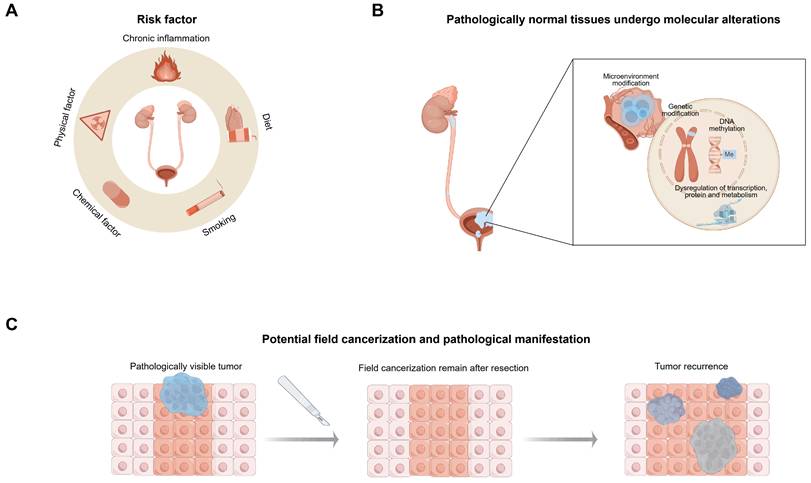
FC refers to molecular alterations that accumulate in tissue after prolonged exposure to carcinogens, inflammation, or other chronic insults, before visible morphological abnormalities develop [13]. In the urothelium, such changes may create a biologically altered background that is more susceptible to malignant transformation. Some regions within this altered field may subsequently acquire additional abnormalities and progress toward dysplasia, carcinoma in situ, invasive disease, or clinically detectable recurrence (Figure 1B) [14-16].
Schematic illustration of the initiation and progression of urothelial carcinoma. (A) Chronic inflammation, unhealthy diet, smoking, as well as physical and chemical carcinogens, are major risk factors for urothelial carcinoma. (B) Under the influence of these carcinogenic factors, histologically normal urothelial epithelium may develop a pathologically invisible field effect, characterized by molecular abnormalities such as genetic alterations, DNA methylation, dysregulation of transcription, translation, and metabolic regulation, accompanied by modifications in the tumor microenvironment. (C) As the disease progresses, urothelial tissues may develop pathologically detectable tumors. Even after tumor resection, the field effect persists, and residual abnormal cells may undergo further malignant transformation, potentially leading to tumor occurrence. By Figdraw.
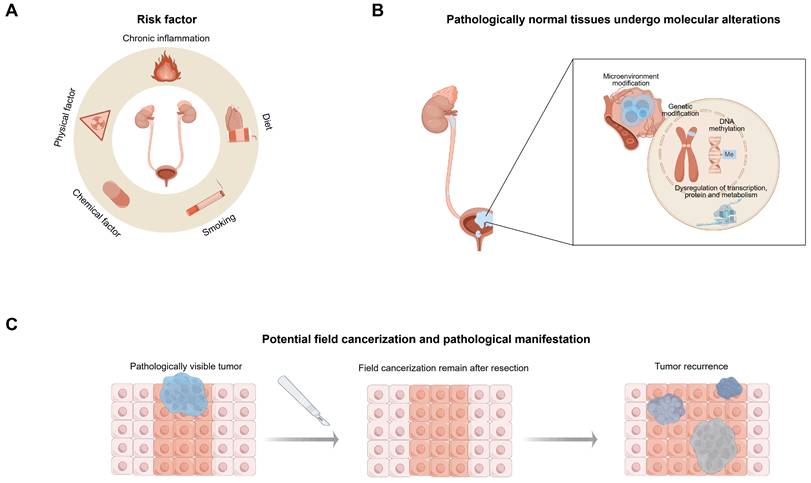
Several established UC risk factors may contribute to this process, including aromatic amines associated with smoking and occupational exposure, arsenic-contaminated drinking water, and recurrent urinary tract infections (Figure 1A) [3]. Long-term exposure to these factors may help explain the multifocal nature and high recurrence rate of UC. Notably, some recurrent lesions show molecular patterns that cannot be readily accounted for by simple clonal spread, providing support for the field cancerization hypothesis [17, 18]. The bladder is particularly suitable for studying this phenomenon because it is the most commonly affected site and allows sampling of adjacent and distant areas of urothelium within the same organ [19].
This review examines the potential contribution of field cancerization to UC initiation, progression, and recurrence. Relevant studies were identified through PubMed and Web of Science searches conducted up to January 2026 using the terms “field cancerization” and “urothelial carcinoma,” followed by manual screening of relevant records.
2. Overview of field cancerization
In 1953, Slaughter and colleagues introduced the concept of field cancerization while studying oral squamous cell carcinoma. They observed that recurrence could not always be explained by the regrowth of the primary tumor alone. Instead, adjacent epithelium often showed gradual proliferative changes and atypical cellular features, suggesting that a broader area of tissue had already been altered before a new lesion became clinically apparent [20]. Under this model, removal of the visible tumor does not necessarily eliminate all biologically abnormal cells. Cells remaining in the surrounding mucosa may continue to accumulate changes and, in some cases, give rise to additional tumor foci or later recurrence (Figure 1C) [21].
The meaning of a “cancerized field” is less clearly standardized in urothelial carcinoma. In most studies, it is inferred when histologically normal-appearing urothelium contains molecular features associated with cancer. Consequently, the boundaries of the field are defined differently across studies. Some investigators compare tissue close to the tumor with samples collected from more distant urothelial sites, asking whether tumor-related alterations extend beyond the grossly visible lesion [22]. Other studies use whole-organ or multi-region sampling to map molecular changes across anatomically documented areas of the bladder mucosa. More recently, spatial profiling and spatial multi-omics approaches have added positional information to these analyses, allowing molecular features to be interpreted in relation to their precise location within the urothelium [16, 23]. Differences in sampling distance, mapping granularity, and analytical platforms therefore introduce inherent challenges when comparing the extent and intensity of field effects across studies.
It is noteworthy that the field cancerization theory does not contradict the traditional theory of clonal expansion. In situ or early recurrence is generally considered to result from the rapid proliferation of the residual tumor clone at the primary site, which is a direct consequence of incomplete surgical and therapeutic eradication, molecularly manifested as monoclonal expansion. In contrast, distant or late recurrence may be promoted by extensive accumulation of field effect, accompanied by subclonal expansion, cooperation among multiple clones, and tumor metastasis [4, 24].
3. Molecular alterations of field cancerization
3.1 Genetic abnormalities
Field cancerization implies that somatic alterations can be detected in histologically normal urothelium. Evidence for such alterations comes from multiple experimental designs with different strengths. Bulk / targeted sequencing of normal biopsies has shown that somatic variants are common even when urothelium appears normal under microscopy. For example, in a targeted panel study of NMIBC, 67% normal urothelium specimens harbored ≥1 somatic mutation, and TERT promoter mutations were detected in 61% of normal samples [25]. A complementary design—deep-targeted sequencing with error correction in selected-site biopsies (SSBs) quantified field effects across multiple bladder locations and time points, and reported that 33% of high-impact mutations in cancer driver genes observed in tumor samples were already present in normal SSBs [15]. Spatial/whole-organ multi-omics mapping provides anatomically resolved evidence that molecular changes can extend beyond visible lesions. Whole-organ histologic and genomic mapping combined bulk RNA-seq, methylation profiling, copy-number variation, and exome mutational profiles to track progression from field effects to carcinoma along luminal and basal trajectories [16]. Copy number and allele-specific analyses (including CN-LOH) capture alterations that point-mutation panels may miss. Using a multi-sample-per-patient protocol and a sensitive pipeline, one cohort reported that 45% of patients had at least one chromosomal alteration in at least one “non-cancerous” urothelium sample, with recurrent hotspots comprising gains/deletions and CN-LOH events [22].
Across studies, recurrent alterations in histologically normal urothelium tend to involve early trunk-like events inferred by shared mutations between normal and tumor, or chromosomal hotspots recurrently observed across patients. In NMIBC targeted sequencing, TERT promoter and FGFR3 alterations were repeatedly observed in normal urothelium, supporting their role as early events compatible with clonal expansion in NMIBC [25]. In the SSB deep-targeted design, 33% of tumor-derived high-impact driver mutations were detectable in normal SSBs, including tumour protein p53 (TP53), AT-rich interactive domain-containing protein 1A, lysine methyltransferase 2D, and phosphatidylinositol-4,5-bisphosphate 3-kinase catalytic subunit alpha [15]. Current studies on the mechanisms of field cancerization indicate that most involved molecules are associated with traditional oncogenes or tumor suppressor genes, but some atypical genes and pathways may also be involved. To explore the key alleles affecting tumor development, Wiktoria Stańkowska et al. analyzed the copy-neutral loss of heterozygosity pattern in the normal urothelium of BC patients, combined with chimeric chromosome amplifications and deletions, and identified several altered gene hotspots. These include genes involved in cell cycle regulation, receptor tyrosine kinases that promote the development of a variety of cancers, and several genes participating in the phosphatidylinositol 3-kinase/protein kinase B, mitogen-activated protein kinase, and wingless/integrase-1 signaling pathways. Many of these genes align with BC, for example, the amplification of E2F transcription factor 3, SRY-related high-mobility-group box 4, fibroblast growth factor receptor 1, and zinc finger protein 703 may be associated with the activation of oncogenes, while deletions of SWI/SNF-related matrix-associated actin-dependent regulator of chromatin subfamily A member 2, nuclear factor I/B, and protein tyrosine phosphatase receptor delta may impair tumor suppression functions. Furthermore, the study also observed some rare genetic variations in BC, such as duplication of fibroblast growth factor receptor 1 and deletion of cyclin-dependent kinase inhibitor 1B, providing new research clues regarding urothelial carcinoma pathogenesis [22]. Notably, the frequency and types of gene mutations in field cancerization show significant heterogeneity across different studies. Some studies suggest that, compared to classical oncogenes, mutations in genes related to chromatin remodeling are more frequent in field cancerization [26, 27]. This phenomenon may be related to the stability of chromatin remodeling genes in tumor development, while mutations in classical oncogenes such as TP53 exhibit more pronounced temporal variation [28].
Importantly, functional evidence in adjacent normal urothelium is usually indirect, most studies do not perform perturbation experiments in normal tissue. Instead, they infer the biological relevance of these alterations by correlating known tumor-suppressor or oncogene functions in bladder cancer with their presence in both normal and tumor compartments. Shared alterations between adjacent normal urothelium and tumor tissue are consistent with clonal continuity and may indicate that some mutations occur early during tumor development. Their relationship with clinical features, including recurrence, progression, or prognosis, may provide further indirect support for biological relevance. Nevertheless, most evidence from adjacent normal tissue remains observational. For example, TP53 inactivation is strongly linked to aggressive bladder cancer, but studies of morphologically normal urothelium generally demonstrate mutation presence or clonal relatedness rather than direct functional effects in experimental models [15, 29].
Molecular changes associated with field cancerization are not uniform across patients, anatomical sites, or molecular subtypes. Different altered urothelial regions may follow distinct evolutionary trajectories, which may contribute to both inter-patient variation and heterogeneity among tumors arising within the same patient [25, 27, 30-33]. Prior radiation exposure may also influence the mutational landscape in a subset of bladder cancers. In radiation-associated cases, alterations in KDM6A and ATM, together with PPARG amplification, have been reported more frequently, whereas CDKN2A mutations appear more common in tumors without a history of radiation exposure [34]. These findings indicate an association between radiation history and particular genomic patterns, but they do not establish that radiation directly produced the observed mutations or the more aggressive clinicopathological features reported in some cohorts.
Mutation frequencies and reported gene lists also differ considerably between studies. Such variation may reflect differences in patient selection, disease stage, tissue sampling, sequencing depth, and analytical strategy. In particular, some deep-targeted sequencing studies examined only mutations already identified in the matched tumor. This approach can increase the apparent proportion of shared mutations while making it harder to detect additional field-associated alterations that were not present in the index tumor [15].
3.2 Epigenetic changes
Field cancerization is not solely driven by genetic mutations; epigenetic regulation also plays an important role. This includes DNA methylation, histone modifications, and chromatin remodeling, among other mechanisms [35, 36]. In urothelial carcinoma, DNA methylation has received particular attention. One practical reason is that methylation changes can be detected not only in tissue samples but also in urine-derived DNA, which makes it possible to explore non-invasive biomarkers and to follow molecular changes over time in a longitudinal manner [37, 38]. DNA methylation primarily occurs in CpG islands within gene promoter regions and is a chemical modification involving the addition of a methyl group to the 5' carbon of cytosine residues. By changing the structure of DNA, methylation can inhibit gene transcription [39].
In samples from patients with urothelial carcinoma, non-cancerous tissues often exhibit detectable DNA methylation changes that are more evident than genetic alterations. Therefore, alterations in DNA methylation may play a more critical role in the field cancerization of the urothelium [40]. A meta-analysis showed that in BC tissues and adjacent normal tissues, the promoter of the death-associated protein kinase (DAPK) gene, located on chromosome 9p34 and involved in the p53-dependent apoptosis pathway, was methylated at similar frequencies, and this was significantly associated with the occurrence of BC [41]. Similarly, in bladder tissues from patients with neurogenic lower urinary tract dysfunction, varying degrees of DNA methylation have been observed in the promoters of tumor-related genes, including Ras association domain family 1, retinoic acid receptors beta, DAPK, human telomerase reverse transcriptase, and adenomatous polyposis coli. This methylation is closely associated with the increased risk of recurrent urinary tract infections and further supports the presence of the epigenetic field effect, suggesting potential risk for BC development [42]. These hypermethylated genes typically lead to the inactivation of tumor suppressor genes, allowing tumor cells to escape normal cellular regulation, and thereby promoting cancer development [43]. However, it is noteworthy that some genes, such as distal-less homeobox 6 and specificity protein 8, exhibit hypomethylation in the field effect. These genes are often related to embryonic development, and tumor cells may activate developmental pathways to enhance their survival and proliferation [16]. In NMIBC, the presence of hypomethylation, unlike in invasive tumors, may provide a biological explanation for its non-invasive behavior [44].
Tadeusz Majewski et al. proposed that widespread mucosal methylation changes may represent an early field alteration for bladder cancer. It suppresses immune responses, and disrupts Ras signaling pathways, leading to the enhanced growth and spread of mutated cells within normal mucosa. Furthermore, signaling pathways affected by DNA copy changes and mutations, such as the Kras pathway, precede the onset of DNA methylation, suggesting that they may enhance mechanisms initiated by methylation [23]. Sangkyou Lee et al. developed a method combining histology and molecular mapping to resolve the initial clonal expansion sequence prior to tumorigenesis. They find that, prior to the loss of tumor suppressor gene function, five genes associated with the field effect of BC located within a 5 Mb region surrounding the retinoblastoma susceptibility gene were downregulated. Further analysis of the lysophosphatidic acid receptor 6 and calcium-binding protein 39-like involved in the luminal and basal subtypes of BC respectively, shows that these genes are silenced by hypermethylation of the CpG site in the promoter, preventing transcription factor binding, affecting cholesterol metabolism and the unfolded protein response, leading to dysregulation of uroepithelial differentiation and promoting cancer progression [45]. However, whether methylation is a true initiating event remains unresolved because most available human data are cross-sectional and observational. Thus, current evidence should be interpreted as suggesting early epigenetic involvement and highlighting the need for longitudinal sampling and functional perturbation studies.
Reported methylation alterations in field cancerization are assay dependent, because targeted MSP/qMSP prioritize predefined loci and maximize analytical sensitivity in low-input urine DNA, whereas Illumina HM450/EPIC arrays and bisulfite-based sequencing (e.g., WGBS) profile broader CpG sets with different coverage and technical biases. As a result, cross-study comparisons and consensus marker selection can be confounded by platform- and workflow-specific effects [46].
Beyond DNA methylation, histone modification and chromatin organization may also contribute to field cancerization. KMT2C and KMT2D are of particular interest because they are frequently inactivated in urothelial carcinoma and, in some studies, in histologically normal-appearing urothelium. These genes encode components of the H3K4me1 methyltransferase machinery.
When KMT2C or KMT2D function is lost, enhancer regulation can be disrupted. This may weaken transcriptional programs involved in urothelial differentiation and is associated with reduced luminal features and expansion of basal-like cell populations. Altered KMT2C/KMT2D activity may also affect the distribution of the KMT2A-menin complex. Rather than remaining at shared enhancers, this complex can become enriched at promoters with bivalent chromatin marks, including H3K4me3 and H3K27me3.
Such redistribution may relieve repression of genes that are normally tightly controlled, particularly immediate early genes and genes involved in inflammatory signaling. In this setting, urothelial cells may become less differentiated and more responsive to growth-related or inflammatory cues. These findings suggest that chromatin remodeling may not directly initiate tumor formation by itself, but could leave the urothelium in a more permissive state for later genetic alterations or continued carcinogen exposure [47].
3.3 Dysregulation of transcription, protein expression, and metabolism
Transcriptomic studies have identified clear molecular differences between urothelial carcinoma subtypes, and these differences may also be relevant to the evolution of field changes [16, 48]. Basal-type bladder cancers are more often associated with invasive disease and commonly express keratin 5 and keratin 6. These tumors also show stronger activation of epithelial-mesenchymal transition (EMT)-related programs. TGF-β1 can promote EMT, whereas p53 has been reported to restrain this process. Transcription factors from the SNAIL, TWIST, ZEB, and FOX families participate in this shift by reducing epithelial markers such as E-cadherin, claudin-1, and tight junction protein 1. TP63 is also involved in the regulation of basal keratins and EMT-associated transcriptional programs in urothelial cells. By comparison, luminal tumors, which are more commonly linked to NMIBC, generally show weaker EMT activity and higher expression of GATA3 [16]. These transcriptional patterns are relevant clinically. Basal programs are usually associated with greater invasiveness and more aggressive biological behavior, whereas luminal programs are more closely related to urothelial differentiation and may show different treatment responses [49].
Proteomic and multi-omics studies further indicate that metabolic regulation, immune activity, and protein homeostasis differ across UC subtypes and disease stages. In one analysis comparing papillary urothelial carcinoma (PUC) with carcinoma in situ (CIS), PUC showed increased FOXO1 and AKT activity, with downstream effects on genes involved in glucose metabolism. CIS, in contrast, showed marked upregulation of JUN and enrichment of pathways related to EMT and immune regulation. Increased infiltration of CD8⁺ T cells, dendritic cells, and M1 macrophages was also observed in CIS.
Metabolic features differed between the two trajectories. In PUC, increased glycolysis and lactate production were negatively associated with immune scores, raising the possibility that lactate-rich conditions may contribute to local immune suppression. During CIS progression, pathways involved in reactive oxygen species metabolism were reduced, while DNA damage-response pathways were increased. This combination may reflect persistent oxidative and genotoxic stress, which could favor genomic instability during progression [50]. The proteogenomic distinction between PUC-derived and CIS-derived trajectories has potential clinical relevance because it maps molecular programs onto progression risk. Another study reported that NMIBC tissues upregulate stress-adaptive and metabolic support proteins such as peroxiredoxin 1, glutathione S-transferase mu 1, and heat shock protein beta-1, whereas MIBC shows prominent increases in a different set of redox homeostasis regulators, including peroxiredoxin 2, biliverdin reductase B, and 15-hydroxyprostaglandin dehydrogenase. This pattern points to stage-dependent changes in antioxidant responses rather than a single, uniform redox program across UC progression [51] Czerniak and colleagues examined protein expression and metabolic alterations in mucosa adjacent to bladder dysplasia and carcinoma in situ [52]. Their findings suggest that molecular disruption is already present before invasive carcinoma becomes apparent. Several pathways involved in energy production, including oxidative phosphorylation, branched-chain amino acid degradation, the tricarboxylic acid cycle, and thermogenesis, were reduced. Together, these changes are consistent with impaired mitochondrial and metabolic capacity.
The study also identified abnormalities in nucleocytoplasmic transport, spliceosome function, ribosome biogenesis, and peroxisomal activity. These findings indicate that protein synthesis, processing, and turnover may already be disturbed in the altered mucosa. Changes in these pathways were linked to dysregulation of peroxisome proliferator-activated receptor signaling, which is relevant to urothelial differentiation, lipid handling, and protein degradation.
Metabolomic data further showed alterations in cysteine and methionine metabolism, fatty acid metabolism, and glycolytic activity. Rather than representing isolated metabolic events, these changes may reflect a broader disturbance in glucose and lipid utilization within the local mucosal environment. Such an altered metabolic background could contribute to the field effect and make the urothelium more permissive to subsequent malignant transformation.
Notably, whole-organ spatial proteomic and metabolomic mapping identified increased lactate levels and enhanced glycolytic activity in microscopically normal-appearing mucosa adjacent to dysplasia or carcinoma in situ. These changes were detectable before overt carcinoma developed. This observation suggests that a Warburg-like metabolic shift may emerge at an early stage of urothelial field cancerization and provides a potentially testable link between metabolic remodeling and later tumor development.
3.4 Microenvironmental Changes
During tumorigenesis and progression, although molecular genetic alterations are extensively studied, the role of the tumor microenvironment is equally critical. The tumor microenvironment consists of various components, including non-cellular elements such as extracellular matrix, bioactive factors, and extracellular vesicles, as well as stromal cells such as fibroblasts, adipocytes, endothelial cells, and immune cells. These factors collectively influence tumor cell survival, invasion, and metastasis [53-55]. In the absence of microenvironmental signals such as inflammation, clonal expansion is unlikely to occur even when mutations are present [56].
The immune-inflammatory response plays a significant role in field carcinogenesis. Jolanta Bondaruk et al. found that basal BC exhibits several mutated genes associated with the inflammatory response, such as the toll-like receptor 4/5, which recognize pathogens and activate innate immune responses, as well as phosphoinositide-3-kinase adapter protein 1, which is involved in the survival of mature B cells and negatively regulates the production of inflammatory cytokines. At the same time, widespread dysregulation of interleukin-related pathways persists during the progression from normal urothelium to urothelial carcinoma [16]. Moreover, in the ostensibly normal mucosal stroma of bladder cancer patients, the numbers of stellate cells and mast cells are reduced. These stromal cells contribute to local immune regulation. Their reduced abundance in apparently normal mucosa may weaken antitumor surveillance and favor a more immunosuppressive environment [57]. The field effect also involves adaptive immune pathways. Basal bladder cancer exhibits mutations in zeta-chain associated protein kinase 70 and POU domain class 2-associating factor 1, which are major regulators of T- and B-cell development, revealing microenvironmental effects on immune cell function and development [16]. Strandgaard et al. assessed CD8⁺ T-cell exhaustion using a transcriptomic score based on the expression of immunoinhibitory genes in tumor RNA-seq data. CD8⁺ T-cell infiltration was estimated computationally. Their results suggested that persistent antigen exposure may contribute to progressive T-cell exhaustion during field carcinogenesis, which could reduce antitumor immune activity and favor tumor progression [15] Overall, tissue-based studies point to an altered local immune environment that may be more permissive for tumor development. Experimental studies of extracellular vesicles (EVs) provide possible mechanisms through which tumor cells may influence adjacent urothelium.
EVs released by bladder cancer cells have been reported to induce lasting changes in susceptible urothelial cells and, under experimental conditions, may promote malignant transformation [58]. Electronic-cigarette exposure can increase EV release from bladder cancer cells. These EVs have been associated with oxidative stress, inflammatory signaling, DNA damage, and increased proliferation in non-malignant urothelial cells [59] Several molecular pathways may be involved. EV-mediated activation of the ERK pathway has been linked to increased expression of CREPT, a transcription-associated protein that can promote chromatin looping and cell-cycle-related transcription. Transcriptomic analyses also suggest that EV exposure activates TNF-related inflammatory signaling. CREPT may further influence this response by regulating TNFR2 and PI3K expression, potentially connecting chronic inflammation with pro-tumorigenic changes in recipient cells [60] The protein cargo carried by tumor-derived EVs may also contribute to these effects. PDIA1, an endoplasmic reticulum oxidoreductase and chaperone protein, has been identified as a potentially important EV component in bladder cancer models. Under endoplasmic reticulum stress, tumor cells may selectively release oxidized and active PDIA1 through EVs. Although this export may reduce intracellular oxidative stress in the donor tumor cells, PDIA1-containing EVs can have different effects after uptake by normal urothelial cells. Experimental evidence indicates that they may induce sustained oxidative stress and DNA damage in recipient cells. Repeated exposure has been associated with malignant transformation and anchorage-independent growth in vitro [61]. Morphologically, exposure to tumor-derived EVs induces significant changes in urothelial cells, including cytoplasmic enlargement, disrupted endoplasmic reticulum (ER) organization, and mitochondrial accumulation. These changes trigger the unfolded protein response in the ER (UPRER). Prolonged UPRER signaling activates survival branches, leading to significant upregulation of inositol-requiring enzyme 1, nuclear factor-kappaB, and the inflammatory cytokines, ultimately suppressing the expression of the pro-apoptotic protein C/EBP homologous protein. These alterations increase genomic instability, disrupt contact inhibition, and enhance cellular invasiveness, creating conditions favorable for tumor progression and dissemination [58]. Taken together, EV-driven oxidative/ER-stress phenotypes provide testable mechanisms for field carcinogenesis, but the strength of evidence currently differs markedly between mechanistic model systems and clinical association studies.
EV-associated signals are attractive for translation in bladder cancer because urine is readily accessible and repeatedly sampled. However, urinary EVs are heterogeneous and can originate from the kidney or upper tract and non-malignant urothelium, potentially diluting bladder-tumor-derived signals. Thus, study design should consider specimen type when “functional” EV biomarkers are pursued [62]. Consistent with this, recent studies and reviews summarize candidate urinary EV cargo classes—including miRNAs, proteins, and glycan/glycoprotein features—for diagnosis, classification, or monitoring of bladder cancer [63]. Yet, reproducibility is limited by variability in EV isolation, characterization and pre-analytical factors. Adherence to community reporting standards and transparent EV characterization will be essential [64].
4. Implications of Field Cancerization for Clinical Strategies
In recent years, driven by the rapid advances in high-throughput sequencing and related technologies, researchers have progressed from bulk sequencing at the population level to single-cell omics analyses that reveal cellular heterogeneity, and further to spatial omics that preserve positional information. This progression enables comprehensive profiling of cell populations within FC regions across the genomic, epigenomic, transcriptomic, proteomic, and metabolomic levels. It can help clarify how molecular changes contribute to UC initiation, progression, and interactions with the surrounding microenvironment, with possible implications for earlier detection and more individualized treatment [65, 66]. Multi-timepoint spatial and temporal omics approaches may further extend this work. Unlike single-timepoint maps, these methods may help track changes in cell populations and local signaling over the course of field cancerization. They could also provide insight into cell-state transitions and evolving interaction networks within altered urothelium and its microenvironment [67, 68]. The major molecular alterations reported in UC-related field cancerization are summarized in Table 1.
Molecular alterations associated with field cancerization in urothelial carcinoma
| Molecular Alteration | Evidence type | Sample | Summary | Ref |
|---|---|---|---|---|
| Gene alterations, microenvironmental changes, urinary proteome changes | Human multi-site tissue sequencing and urine biomarker study | High-risk NMIBC treated with BCG: 751 selected-site bladder biopsies (662 normal-appearing urothelium, 79 lesions) from 70 patients; 234 urine samples from 112 patients; 223 tumors from 136 patients. | Error-corrected sequencing identified mutations in normal-appearing urothelium, including KDM6A, ARID1A, and TP53. High field level linked to CD8 T-cell exhaustion and worse post-BCG outcomes; utDNA after BCG predicted recurrence/progression and high field increased VEGFA and CD27. | [15] |
| Gene alterations | Human longitudinal tumor evolution study | Serial TURBT specimens from 73 patients progressing to MIBC, metastasis, or radical cystectomy: 357 FFPE tumors; 271 tumors for immunohistochemistry subtyping; 200 tumors for gene-expression profiling; 220 tumors for hotspot genotyping. | Longitudinal analysis showed that hotspot FGFR3, PIK3CA, and TERT promoter alterations were largely preserved across recurrences, whereas progression toward more aggressive subtypes was associated with abnormal p53 staining. | [29] |
| Gene alterations | Human normal-appearing urothelium sequencing/mapping study | Laser-microdissected histologically normal urothelium from 15 transplant organ donors (1,647 microbiopsies) and 5 bladder cancer patients (450 microbiopsies); targeted 321-gene panel, WES, and WGS. | Patchwork of microscopic clones (median 40 exome/1879 genome mutations); 17 positively selected genes dominated by chromatin remodeling; APOBEC-positive microbiopsies 22%; TP53 rare, FGFR3 and TERT promoter absent. | [27] |
| Gene alterations | Human tumor genomic association study | 82 bladder tumors analyzed by targeted NGS: 41 with prior pelvic radiotherapy for prostate cancer and 41 without prior radiotherapy. | Recurrent variants showed CDKN2A mutation enriched in non-radiotherapy controls, while ATM enriched in 0-10y post-radiotherapy and KDM6A enriched ≥10y. | [34] |
| Gene alterations | Human normal-appearing margin chromosomal analysis study | Primary tumors, matched histologically normal urothelial margins sampled 1-2 cm (proximal) and 4-5 cm (distal) from the tumor, and whole blood from 67 patients (145 tumors, 277 margins, and 63 blood samples). | SNP-array/MoChA detected low-fraction post-zygotic chromosomal gains, deletions, and copy-neutral loss of heterozygosity in 45% of patients' histologically normal margins; 16 recurrent hotspots overlapped bladder cancer driver regions, supporting field cancerization beyond histology. | [22] |
| Gene alterations | Human normal-appearing urothelium sequencing study | Laser-capture microdissected morphologically normal bladder/ureter urothelium (161 samples from 120 patients), with matched tumors (n = 126) and blood; whole-exome sequencing. | Morphologically normal urothelium contained macroscopic mutant clones, with frequent KMT2D and KDM6A alterations but rare TP53, PIK3CA, and FGFR3 mutations. | [26] |
| Gene alterations | Human targeted sequencing study of normal urothelium and paired NMIBC | TURBT tissues from 90 BCG-naive NMIBC patients (124 tissues) and 34 post-BCG relapse patients; deep targeted sequencing with matched normal. | TERT promoter and chromatin-remodeling alterations were relatively stable, whereas FGFR3, PIK3CA, TSC1, and TP53 changed more dynamically over time. ARID1A mutation was associated with relapse risk, and CCNE1 amplification with progression risk, supporting persistence of altered precursor populations with ongoing evolution after BCG. | [28] |
| Epigenetic alterations | Human methylation study of non-cancerous urothelium | Non-cancerous urothelium from UC patients (N = 47) with paired tumors (T = 46) and control urothelium (C = 26); genome-wide DNA methylation profiling. | 2,502 differentially methylated CpG probes between non-cancerous urothelium and controls. Many of these alterations were retained or strengthened in paired tumors. | [40] |
| Epigenetic alterations | Human methylation study in non-malignant bladder mucosa | Cold-cup bladder biopsies from patients with neurogenic lower urinary tract dysfunction (n = 24); qMSP analysis. | RARB, RASSF1, and DAPK promoter hypermethylation was frequent. | [42] |
| Gene, epigenetic, and transcriptional alterations | Human whole-organ spatial multi-omics mapping study | Normal urothelium, low-/high-grade intraurothelial neoplasia, and UC from 9 cystectomy bladders; Luminal map24 and basal map19 with ureter normal controls and matched blood germline. | Spatial multi-omics identified early field changes across anatomically mapped mucosa, including aberrant CpG methylation, monotonic expression plaques, and expanding clonal mutations/CNVs. | [16] |
| Epigenetic alterations | Human methylation study of matched normal-appearing urothelium | 49 Ta-T1 and 38 T2-T4 UC; matched normal-appearing urothelium microdissected (≥5 cm from invasive tumor); 12 cancer-free urothelium controls. | Ta-T1 tumors were hypomethylated, whereas T2-T4 tumors were broadly hypermethylated; matched normal-appearing urothelium already showed hypermethylation at about 12% of loci, supporting a polyclonal epigenetic field defect consistent with FC. | [44] |
| Gene and epigenetic alterations | Human whole-organ methylation field-mapping study | Single cystectomy bladder mapped into 41 1×2 cm blocks; scraped mucosa or microdissected UC (>90% urothelial/tumor cells); matched blood DNA; methylation controls: ureter urothelium from 3 nephrectomies. | Field-effect mucosa (normal urothelium/low-grade intraurothelial neoplasia) showed early driver mutations (TERT, KDM6A, ARID1A) and widespread CpG-island dysmethylation, with low-level CNVs. | [23] |
| Epigenetic alterations | Human whole-organ promoter methylation/LOP mapping study | Whole-organ cystectomy maps (n=20): bladder mucosa partitioned into 1×2 cm blocks (902 geo-annotated samples; plus primary tumors (n=189) and normal urothelium controls; promoter methylation by quantitative methylation-specific PCR/bisulfite sequencing; LOP around RB1 by SNPlex/SNP allelotyping. | Plaque-like promoter hypermethylation silenced forerunner genes LPAR6 or CAB39L and extended into NU with minimal/no morphologic change, inversely correlating with expression. LPAR6 and CAB39L were methylated in 72% and 92% of tumors, biasing urothelial basal-to-luminal differentiation toward luminal/papillary vs basal states. | [45] |
| Gene and transcriptional alterations | Human longitudinal tumor evolution study | Serial primary and recurrent bladder tumors (18 specimens) from 5 patients, including locoregional and distant metastases, with matched blood; whole-exome sequencing and RNA sequencing. | Recurrent lesions largely retained early clonal driver mutations but acquired additional subclonal expansions, and RNA sequencing indicated switching between basal and luminal expression subtypes over time and sites, consistent with evolving residual populations driving relapse. | [48] |
| Transcriptional and metabolic alterations | Human multi-omics proteogenomic study | FFPE urothelial bladder lesions across stages from 190 UC patients; proteome n=448, phosphoproteome n=211, whole-exome n=125, transcriptome n=67. | Papilloma was dominated by HRAS hotspot mutations (83%) with MAPK activation and mTORC1 pathway dysregulation. PUC showed higher glucolipid metabolism and lower immune infiltration, whereas CIS showed DNA damage response, APOBEC signature, RBPMS loss, and activator protein 1 (AP-1) activation. | [50] |
| Proteomic alterations | Human tissue proteomic comparison study | UBC tumor tissue (NMIBC Ta/T1 n=25; MIBC T2-T4 n=17) and noncancerous bladder tissue (n=10); proteomic profiling. | Twelve proteins differed, 9 higher in NMIBC and 3 higher in MIBC, dominated by oxidative stress and antioxidant functions, including PRDX1/PRDX6 versus PRDX2, biliverdin reductase B, and 15-hydroxyprostaglandin dehydrogenase, suggesting redox adaptation as candidate biomarkers. | [51] |
| Proteomic and metabolic alterations | Human whole-organ spatial proteomic/metabolomic mapping study | Radical cystectomy, whole-organ grid sampling across mucosa and tumor; DNA, proteome, and metabolome from each well; whole-exome sequencing plus LC-MS/MS proteomics and targeted LC-MS/MS metabolomics. | Across normal urothelium, low- and high-grade intraurothelial neoplasia, and invasive UC, protein homeostasis pathways and PPAR-regulated lipid/carbohydrate metabolism were dysregulated; mutations clustered by spread and VAF into alpha, beta (below 20%), and gamma (above 20%) patterns, with gamma expanding. | [52] |
| Microenvironmental alterations | Human tissue immunohistochemistry study | Bladder neoplasia (n=52) and benign bladder conditions (n=18); benign-appearing mucosa and neoplastic tissue; immunohistochemistry for ALDH1A1 and CD44 plus differentiation markers. | Compared with benign controls, benign-appearing mucosa from UC patients showed fewer ALDH1A1-positive stromal stellate cells and mast cells, consistent with stromal field-effect remodeling near cancer. | [57] |
| Microenvironmental alterations via extracellular vesicles | Cell/extracellular vesicle transformation model | SV-HUC urothelial cells treated twice weekly with bladder cancer cell-derived EVs (20 μg/mL, 8-13 weeks; 12 weeks plus 4-week washout), including cigarette-smoke extract (1%) or unflavored/menthol E-liquid-stimulated EVs from TCCSUP/T24; soft agar colonies and athymic nude-mouse xenografts. | EV uptake induced endoplasmic reticulum stress with sustained unfolded protein response signaling (PERK to IRE1 shift, NF-kappaB activation), inflammatory cytokine release, oxidative stress, and DNA damage, leading to contact-inhibition escape, invasion, and anchorage-independent growth, consistent with FC-driven transformation. | [58, 59] |
Field effects are generally viewed as a source of risk because altered urothelial areas may provide a background for multifocal tumor formation. However, mutated clones in normal tissue do not necessarily behave in the same way as tumor clones. In some settings, clonal competition within normal epithelium may remove or suppress early neoplastic cells [69] The expansion of certain mutant populations may therefore reflect a tissue-level selection process rather than inevitable malignant progression. Although this process can increase the number of detectable mutations in apparently healthy tissue, it may occasionally limit the growth of more harmful clones [70]. This complexity should be considered when interpreting molecular abnormalities in cancer-prone fields.
From a translational perspective, field cancerization may offer opportunities for risk assessment, early detection, and follow-up after treatment. Smoking, chronic inflammation or infection, dietary factors, chemical exposure, and physical carcinogenic stimuli are all relevant to the development of altered urothelial fields, which also emphasizes the value of prevention [71, 72]. Clinically, applying optical imaging technologies to develop fluorescent probes that specifically bind early gene mutations or DNA methylation sites could enable precise localization of FC areas and early tumor detection under cystoscopy. Clinical studies have supported that hexaminolevulinate-guided fluorescence cystoscopy can improve detection and reduce early recurrence compared with white light cystoscopy alone, indicating that optical enhancement can be clinically actionable when integrated into standard cystoscopic workflows [73]. Building beyond this precedent, near-term optical probe development is generally more feasible when targeting accessible cell-surface proteins, whereas urine-based mutation and methylation assays can serve as noninvasive tools for risk enrichment to define high-risk patients or fields [74, 75]. In this context, candidate targets with translational evidence include CD47-directed fluorescence imaging in human bladder tissues and a CD44v6-targeted near infrared fluorescent agent evaluated in a first-in-human endoscopic study [76, 77]. In addition, clinical translation of targeted optical probes will require prospective evaluation of safety and diagnostic accuracy within standardized imaging protocols and an appropriate regulatory framework for agent-based imaging during cystoscopy. Targeting microenvironmental changes may facilitate novel immunotherapeutic agents to reduce tumor recurrence [5, 78-80]. However, due to the limited availability of clinical specimens, incorporating FC research into animal models is essential. By performing multi-site, longitudinal sampling to dynamically analyze molecular evolution from normal through precancerous to tumor stages, we can optimize model systems that recapitulate human lesion progression, facilitating the development of urothelial carcinoma animal models that closely resemble human cancer progression and support clinical drug research [70].
Recent studies indicate that urinary tumor DNA (utDNA) and circulating tumor DNA (ctDNA) hold promise as prospective tools for detecting minimal residual disease and metastatic recurrence. Both approaches may detect molecular residual disease more sensitively than conventional pathological assessment. Detectable utDNA or ctDNA may originate from overt tumor cells, occult microscopic disease, or molecularly altered cells within histologically normal-appearing urothelium. Higher utDNA and ctDNA levels have also been associated with unfavorable clinical outcomes and an increased risk of recurrence [15, 81-83] These assays may provide additional information during surveillance and could eventually reduce reliance on frequent cystoscopy in selected patients. Their limitations should nevertheless be recognized. A negative liquid-biopsy result can be difficult to interpret when tumor-derived DNA is present at very low levels, as assay sensitivity declines with low analyte abundance [84]. Plasma ctDNA testing may also be affected by clonal hematopoiesis, which can introduce biological noise and false-positive signals. Matched leukocyte sequencing or appropriate filtering methods may help address this issue [85] In a cohort of patients undergoing radical cystectomy with curative intent, utDNA-based minimal residual disease detection predicted pathological response with 81% sensitivity and 81% specificity [86]. These results support the clinical potential of utDNA testing, although a negative result should not be considered definitive evidence that residual disease is absent.
5. Current challenges and future prospects
Field cancerization has broadened the understanding of urothelial carcinoma development, but several practical and conceptual issues remain unresolved. Spatial and temporal transcriptomic methods can map cell populations and transcriptional programs within tissue, yet their broader use is still limited by cost, technical demands, and the scale of data processing required. Many available studies are based on cross-sectional samples and use computational trajectory analysis to infer cell-state transitions [67]. Such pseudotime analyses are useful, but they cannot replace prospective longitudinal sampling. Large datasets that include repeated sampling and precise anatomical information are still scarce [87]. Future studies will need designs that balance molecular depth with feasible cohort sizes. When possible, whole-organ mapping or geographically documented sampling should be incorporated to clarify how far field-associated alterations extend within the urothelium [23].
Tissue collection presents another challenge. Repeated biopsies from several sites may increase patient burden and raise ethical concerns, particularly when samples are taken from areas that appear normal on routine pathology. Small differences in sampling location can also affect molecular findings and make comparisons between studies difficult [88, 89]. One practical solution may be the use of standardized biobanking procedures. Preserving residual mucosa from surgical specimens, together with detailed information on sampling location, could reduce the need for additional research biopsies and improve reproducibility across cohorts. It is also important to note that most current evidence comes from bladder cancer. Direct evidence for field cancerization in upper tract urothelial carcinoma remains limited, and findings from bladder-based studies should not be automatically applied to upper tract disease without further validation.
Experimental models also have limitations. Cell culture systems and conventional animal models cannot fully reproduce the complex epithelial, stromal, immune, and spatial interactions present in the human urothelial field [90-93]. Animal studies should therefore be interpreted as models of early mucosal alteration rather than exact representations of human field cancerization. For example, introducing Trp53 mutations into Krt5-expressing basal cells increased the incidence of muscle-invasive bladder cancer in a BBN-induced model, indicating that lineage-specific genetic changes can interact with chronic carcinogen exposure to influence disease progression [94].
A further issue is the lack of a widely accepted definition of field cancerization in urothelial carcinoma. The concept was initially developed in head and neck cancer research, and its application to urothelial tumors remains inconsistent. Studies often focus on molecular abnormalities in tissue adjacent to tumors but do not always address the wider anatomical distribution or clinical relevance of these changes [95, 96]. Future work would benefit from reporting sampling sites in a standardized and geographically explicit manner. Important analytical parameters should also be predefined and reported clearly. Where appropriate, data should be deposited in controlled-access repositories so that findings can be independently examined while protecting patient privacy.
Progress in this area will likely depend on three related developments. First, a practical definition of field cancerization is needed that combines histological appearance with anatomical location and molecular features. Second, prospective studies with geographically documented sampling are required to improve comparability between cohorts. Third, translational approaches should be tested in clinical settings, particularly urine-based molecular assays, imaging methods, and prevention-oriented interventions. The priority should not simply be to generate more descriptive data, but to determine whether field-associated molecular changes can improve patient stratification, surveillance, or treatment decisions.
6. Conclusions
Current evidence indicates that field cancerization is a relevant biological feature of urothelial carcinogenesis rather than only a descriptive observation. Genetic, epigenetic, metabolic, and microenvironmental abnormalities can be detected in urothelium that appears normal by conventional histology, suggesting that molecular changes may arise before an overt lesion is visible.
Field cancerization does not replace the clonal expansion model. Instead, it offers an additional explanation for multifocal disease, recurrence, and molecular heterogeneity between urothelial lesions. Its potential clinical value lies mainly in molecular risk assessment and in the development of surveillance tools based on urine testing or imaging. However, routine application will require standardized definitions, consistent sampling strategies, and prospective validation in clinically representative cohorts.
Overall, field cancerization provides a useful framework for understanding how altered urothelial tissue may contribute to tumor development and recurrence. Further work should focus on translating these observations into approaches that support earlier detection, more accurate risk assessment, and better prevention of recurrence.
Acknowledgements
Funding
This study was supported by the National Natural Science Foundation of China (grant number 82160583), and the Graduate Innovation Fund project of Kunming Medical University: 2025S092.
Author Contributions
X.Y. conceived the review and drafted the manuscript. J.C., H.L., H.D., T.L., and P.C. helped revise this manuscript; S.F. and H.W. designed this review concept and helped edit and revise the manuscript. All the authors read and approved the manuscript.
Artificial Intelligence (AI) usage statement
During manuscript preparation, ChatGPT was used for English-language polishing, including grammar correction, improvement of clarity, and refinement of academic expression.
Competing Interests
The authors have declared that no competing interest exists.
References
1. Yip W, Jaime-Casas S, Kothari A. et al. Urothelial carcinoma: Perioperative considerations from top to bottom. CA Cancer J Clin. 2025;75:528-551
2. Dyrskjøt L, Hansel DE, Efstathiou JA. et al. Bladder cancer. Nat Rev Dis Primers. 2023;9:58
3. Jubber I, Ong S, Bukavina L. et al. Epidemiology of Bladder Cancer in 2023: A Systematic Review of Risk Factors. Eur Urol. 2023;84:176-190
4. Teoh JY, Kamat AM, Black PC. et al. Recurrence mechanisms of non-muscle-invasive bladder cancer - a clinical perspective. Nat Rev Urol. 2022;19:280-294
5. Lopez-Beltran A, Cookson MS, Guercio BJ. et al. Advances in diagnosis and treatment of bladder cancer. BMJ. 2024;384:e76743
6. Paner GP, Smith SC, Hartmann A. et al. Flat intraurothelial lesions of the urinary bladder-do hyperplasia, dysplasia, and atypia of unknown significance need to exist as diagnostic entities? and how to handle in routine clinical practice. Mod Pathol. 2022;35:1296-1305
7. van Doeveren T, Nakauma Gonzalez JA, Mason AS. et al. The clonal relation of primary upper urinary tract urothelial carcinoma and paired urothelial carcinoma of the bladder. Int J Cancer. 2021;148:981-987
8. Gu X, Wang L, Coates PJ. et al. Evidence for etiologic field changes in tongue distant from tumor in patients with squamous cell carcinoma of the oral tongue. J Pathol. 2023;259:93-102
9. Kadara H, Sivakumar S, Jakubek Y. et al. Driver Mutations in Normal Airway Epithelium Elucidate Spatiotemporal Resolution of Lung Cancer. Am J Respir Crit Care Med. 2019;200:742-750
10. Yokoyama A, Kakiuchi N, Yoshizato T. et al. Age-related remodelling of oesophageal epithelia by mutated cancer drivers. Nature. 2019;565:312-317
11. Ikuta S, Saito Y, Takata S. et al. Variability in non-tumor areas of colorectal cancer patients as revealed by endoscopic intestinal step biopsies. Mol Cancer. 2024;23:249
12. Nishimura T, Kakiuchi N, Yoshida K. et al. Evolutionary histories of breast cancer and related clones. Nature. 2023;620:607-614
13. Rahal Z, Sinjab A, Wistuba II. et al. Game of clones: Battles in the field of carcinogenesis. Pharmacol Ther. 2022;237:108251
14. van Doeveren T, van de Werken HJG, van Riet J. et al. Synchronous and metachronous urothelial carcinoma of the upper urinary tract and the bladder: Are they clonally related? A systematic review. Urol Oncol. 2020;38:590-598
15. Strandgaard T, Nordentoft I, Birkenkamp-Demtröder K. et al. Field Cancerization Is Associated with Tumor Development, T-cell Exhaustion, and Clinical Outcomes in Bladder Cancer. Eur Urol. 2024;85:82-92
16. Bondaruk J, Jaksik R, Wang Z. et al. The origin of bladder cancer from mucosal field effects. iScience. 2022;25:104551
17. Mertens LS, Sharma V, Matin SF. et al. Bladder Recurrence Following Upper Tract Surgery for Urothelial Carcinoma: A Contemporary Review of Risk Factors and Management Strategies. Eur Urol Open Sci. 2023;49:60-66
18. Shigeta K, Matsumoto K, Tanaka N. et al. Profiling the Biological Characteristics and Transitions through Upper Tract Tumor Origin, Bladder Recurrence, and Muscle-Invasive Bladder Progression in Upper Tract Urothelial Carcinoma. Int J Mol Sci. 2022;23:5154
19. Czerniak B, Dinney C, McConkey D. Origins of Bladder Cancer. Annu Rev Pathol. 2016;11:149-174
20. Slaughter DP, Southwick HW, Smejkal W. Field cancerization in oral stratified squamous epithelium; clinical implications of multicentric origin. Cancer. 1953;6:963-968
21. Pierik AS, Leemans CR, Brakenhoff RH. Resection Margins in Head and Neck Cancer Surgery: An Update of Residual Disease and Field Cancerization. Cancers (Basel). 2021;13:2635
22. Stańkowska W, Sarkisyan D, Bruhn-Olszewska B. et al. Tumor Predisposing Post-Zygotic Chromosomal Alterations in Bladder Cancer-Insights from Histologically Normal Urothelium. Cancers (Basel). 2024;16:961
23. Majewski T, Yao H, Bondaruk J. et al. Whole-Organ Genomic Characterization of Mucosal Field Effects Initiating Bladder Carcinogenesis. Cell Rep. 2019;26:2241-2256
24. Dang HX, Krasnick BA, White BS. et al. The clonal evolution of metastatic colorectal cancer. Sci Adv. 2020;6:eaay9691
25. Hayashi Y, Fujita K, Sakai K. et al. Targeted-sequence of normal urothelium and tumor of patients with non-muscle invasive bladder cancer. Sci Rep. 2022;12:16642
26. Li R, Du Y, Chen Z. et al. Macroscopic somatic clonal expansion in morphologically normal human urothelium. Science. 2020;370:82-89
27. Lawson ARJ, Abascal F, Coorens THH. et al. Extensive heterogeneity in somatic mutation and selection in the human bladder. Science. 2020;370:75-82
28. Bacon JVW, Müller DC, Ritch E. et al. Somatic Features of Response and Relapse in Non-muscle-invasive Bladder Cancer Treated with Bacillus Calmette-Guérin Immunotherapy. Eur Urol Oncol. 2022;5:677-686
29. Sjödahl G, Eriksson P, Patschan O. et al. Molecular changes during progression from nonmuscle invasive to advanced urothelial carcinoma. Int J Cancer. 2020;146:2636-2647
30. Park HS, Shin H, Lee M. et al. Seminal Vesicle Involvement by Carcinoma In Situ of the Bladder: Clonal Analysis Using Next-Generation Sequencing to Elucidate the Mechanism of Tumor Spread. Cancer Res Treat. 2020;52:1283-1287
31. Clinton TN, Chen Z, Wise H. et al. Genomic heterogeneity as a barrier to precision oncology in urothelial cancer. Cell Rep. 2022;41:111859
32. Grant SR, Tang L, Wei L. et al. Mutation Hotspots Found in Bladder Cancer Aid Prediction of Carcinogenic Risk in Normal Urothelium. Int J Mol Sci. 2023;24:7852
33. Sirab N, Drubay D, Maillé P. et al. Multilayer spectrum of intratumoral heterogeneity in basal bladder cancer. J Pathol. 2022;256:108-118
34. Wijetunga NA, Gessner KH, Kanchi K. et al. Poor Prognosis among Radiation-Associated Bladder Cancer Is Defined by Clinicogenomic Features. Cancer Res Commun. 2024;4:2320-2334
35. Hussain S, Tulsyan S, Dar SA. et al. Role of epigenetics in carcinogenesis: Recent advancements in anticancer therapy. Semin Cancer Biol. 2022;83:441-451
36. Chen X, Xie R, Gu P. et al. Long Noncoding RNA LBCS Inhibits Self-Renewal and Chemoresistance of Bladder Cancer Stem Cells through Epigenetic Silencing of SOX2. Clin Cancer Res. 2019;25:1389-1403
37. Nunes SP, Henrique R, Jerónimo C. et al. DNA Methylation as a Therapeutic Target for Bladder Cancer. Cells. 2020;9:1850
38. Silva-Ferreira M, Carvalho JA, Salta S. et al. Diagnostic Test Accuracy of Urinary DNA Methylation-based Biomarkers for the Detection of Primary and Recurrent Bladder Cancer: A Systematic Review and Meta-analysis. Eur Urol Focus. 2024;10:922-934
39. Soto-Palma C, Niedernhofer LJ, Faulk CD. et al. Epigenetics, DNA damage, and aging. J Clin Invest. 2022;132:e158446
40. Tsumura K, Arai E, Tian Y. et al. Establishment of permutation for cancer risk estimation in the urothelium based on genome-wide DNA methylation analysis. Carcinogenesis. 2019;40:1308-1319
41. Dai L, Ma C, Zhang Z. et al. DAPK Promoter Methylation and Bladder Cancer Risk: A Systematic Review and Meta-Analysis. PLoS One. 2016;11:e167228
42. Koukourikis P, Papaioannou M, Pervana S. et al. Exploring the DNA Methylation Profile of Genes Associated with Bladder Cancer in Bladder Tissue of Patients with Neurogenic Lower Urinary Tract Dysfunction. Int J Mol Sci. 2024;25:5660
43. Nishiyama A, Nakanishi M. Navigating the DNA methylation landscape of cancer. Trends Genet. 2021;37:1012-1027
44. Wolff EM, Chihara Y, Pan F. et al. Unique DNA Methylation Patterns Distinguish Noninvasive and Invasive Urothelial Cancers and Establish an Epigenetic Field Defect in Premalignant Tissue. Cancer Res. 2010;70:8169-8178
45. Lee S, Bondaruk J, Wang Y. et al. Loss of LPAR6 and CAB39L dysregulates the basal-to-luminal urothelial differentiation program, contributing to bladder carcinogenesis. Cell Rep. 2024;43:114146
46. de Abreu AR, Ibrahim J, Lemonidis V. et al. Comparison of current methods for genome-wide DNA methylation profiling. Epigenetics Chromatin. 2025;18:57
47. Wang N, Pachai MR, Li D. et al. Loss of Kmt2c or Kmt2d primes urothelium for tumorigenesis and redistributes KMT2A-menin to bivalent promoters. Nat Genet. 2025;57:165-179
48. Ryu D, Kim T, Lee Y. et al. Longitudinal Analyses of Mutational Subclonal Architecture and Tumor Subtypes in Recurrent Bladder Cancer. Int J Mol Sci. 2023;24:8418
49. Choi W, Porten S, Kim S. et al. Identification of distinct basal and luminal subtypes of muscle-invasive bladder cancer with different sensitivities to frontline chemotherapy. Cancer Cell. 2014;25:152-165
50. Yao Z, Xu N, Shang G. et al. Proteogenomics of different urothelial bladder cancer stages reveals distinct molecular features for papillary cancer and carcinoma in situ. Nat Commun. 2023;14:5670
51. Tabaei S, Haghshenas MR, Ariafar A. et al. Comparative proteomics analysis in different stages of urothelial bladder cancer for identification of potential biomarkers: highlighted role for antioxidant activity. Clin Proteomics. 2023;20:28
52. Czerniak B, Lee S, Jung SY. et al. Inferring Bladder Cancer Evolution from Mucosal Field Effects by Whole-Organ Spatial Mutational, Proteomic, and Metabolomic Mapping. Res Sq [Preprint]. 2024 rs.3.rs-3994376
53. Eguchi T, Sheta M, Fujii M. et al. Cancer extracellular vesicles, tumoroid models, and tumor microenvironment. Semin Cancer Biol. 2022;86(Pt 1):112-126
54. Yuan S, Almagro J, Fuchs E. Beyond genetics: driving cancer with the tumour microenvironment behind the wheel. Nat Rev Cancer. 2024;24:274-286
55. Zhang Q, Liu S, Wang H. et al. ETV4 Mediated Tumor-Associated Neutrophil Infiltration Facilitates Lymphangiogenesis and Lymphatic Metastasis of Bladder Cancer. Adv Sci (Weinh). 2023;10:e2205613
56. Sinjab A, Han G, Wang L. et al. Field Carcinogenesis in Cancer Evolution: What the Cell Is Going On? Cancer Res. 2020;80:4888-4891
57. Isfoss BL, Busch C, Hermelin H. et al. Stem cell marker-positive stellate cells and mast cells are reduced in benign-appearing bladder tissue in patients with urothelial carcinoma. Virchows Arch. 2014;464:473-488
58. Wu C, Silvers CR, Messing EM. et al. Bladder cancer extracellular vesicles drive tumorigenesis by inducing the unfolded protein response in endoplasmic reticulum of nonmalignant cells. J Biol Chem. 2019;294:3207-3218
59. Molony RD, Wu C, Lee Y. E-liquid exposure induces bladder cancer cells to release extracellular vesicles that promote non-malignant urothelial cell transformation. Sci Rep. 2023;13:142
60. Lin Y, Jiang H, Li J. et al. Microenvironment-induced CREPT expression by cancer-derived small extracellular vesicles primes field cancerization. Theranostics. 2024;14:662-680
61. Wu C, Yuen KL, Molony RD. et al. Protein disulfide isomerase-enriched extracellular vesicles from bladder cancer cells support tumor survival and malignant transformation in the bladder. Oncogene. 2025;44:2158-2169
62. Tomiyama E, Matsuzaki K, Fujita K. et al. Proteomic analysis of urinary and tissue-exudative extracellular vesicles to discover novel bladder cancer biomarkers. Cancer Sci. 2021;112:2033-2045
63. Kural S, Jain G, Agarwal S. et al. Urinary extracellular vesicles-encapsulated miRNA signatures: A new paradigm for urinary bladder cancer diagnosis and classification. Urol Oncol. 2024;42:179-190
64. Welsh JA, Goberdhan DCI, O'Driscoll L. et al. Minimal information for studies of extracellular vesicles (MISEV2023): From basic to advanced approaches. J Extracell Vesicles. 2024;13:e12404
65. Akhoundova D, Rubin MA. Clinical application of advanced multi-omics tumor profiling: Shaping precision oncology of the future. Cancer Cell. 2022;40:920-938
66. Parris BA, Shaw E, Pang B. et al. Somatic mutations and immune checkpoint biomarkers. Respirology. 2019;24:215-226
67. Liu L, Chen A, Li Y. et al. Spatiotemporal omics for biology and medicine. Cell. 2024;187:4488-4519
68. Lv W, Zeng Y, Li C. et al. Spatial-Temporal Diversity of Extrachromosomal DNA Shapes Urothelial Carcinoma Evolution and the Tumor Immune Microenvironment. Cancer Discov. 2025;15:1225-1246
69. Colom B, Herms A, Hall MWJ. et al. Mutant clones in normal epithelium outcompete and eliminate emerging tumours. Nature. 2021;598:510-514
70. Ciwinska M, Messal HA, Hristova HR. et al. Mechanisms that clear mutations drive field cancerization in mammary tissue. Nature. 2024;633:198-206
71. Lochhead P, Chan AT, Nishihara R. et al. Etiologic field effect: reappraisal of the field effect concept in cancer predisposition and progression. Mod Pathol. 2015;28:14-29
72. Curtius K, Wright NA, Graham TA. An evolutionary perspective on field cancerization. Nat Rev Cancer. 2018;18:19-32
73. Pohar KS. Blue Light Cystoscopy: Indications and Outcomes. Curr Urol Rep. 2020;21:19
74. Chen X, Zhang J, Ruan W. et al. Urine DNA methylation assay enables early detection and recurrence monitoring for bladder cancer. J Clin Invest. 2020;130:6278-6289
75. Ruan W, Chen X, Huang M. et al. A urine-based DNA methylation assay to facilitate early detection and risk stratification of bladder cancer. Clin Epigenetics. 2021;13:91
76. Pan Y, Volkmer J, Mach KE. et al. Endoscopic molecular imaging of human bladder cancer using a CD47 antibody. Sci Transl Med. 2014;6:260ra148
77. Shang W, Peng L, He K. et al. A clinical study of a CD44v6-targeted fluorescent agent for the detection of non-muscle invasive bladder cancer. Eur J Nucl Med Mol Imaging. 2022;49:3033-3045
78. Zhan H, Wang H, Pan B. et al. Reprogramming the tumor microenvironment with c-MYC-based gene circuit platform to enhance specific cancer immunotherapy. Nat Commun. 2025;16:7983
79. Wang H, Zhan H, Pan B. et al. Engineering CRISPR System-Based Bacterial Outer Membrane Vesicle Potentiates T Cell Immunity for Enhanced Cancer Immunotherapy. Adv Mater. 2025;37:e2501565
80. Zhang C, Liu S, Zhang J. et al. A Multifunctional Fe-EGCG@RSL3 Nanomedicine Synergizes Ferroptosis Induction and Tumor Microenvironment Remodeling for Enhanced Bladder Cancer Immunotherapy. Research (Wash D C). 2025;8:735
81. Lindskrog SV, Birkenkamp-Demtröder K, Nordentoft I. et al. Circulating Tumor DNA Analysis in Advanced Urothelial Carcinoma: Insights from Biological Analysis and Extended Clinical Follow-up. Clin Cancer Res. 2023;29:4797-4807
82. Huelster HL, Gould B, Schiftan EA. et al. Novel Use of Circulating Tumor DNA to Identify Muscle-invasive and Non-organ-confined Upper Tract Urothelial Carcinoma. Eur Urol. 2024;85:283-292
83. Vedeld HM, Pharo H, Sørbø AK. et al. Distinct longitudinal patterns of urine tumor DNA in patients undergoing surveillance for bladder cancer. Mol Oncol. 2024;18:2684-2695
84. Semenkovich NP, Szymanski JJ, Earland N. et al. Genomic approaches to cancer and minimal residual disease detection using circulating tumor DNA. J Immunother Cancer. 2023;11:e006284
85. Liu J, Chen X, Wang J. et al. Biological background of the genomic variations of cf-DNA in healthy individuals. Ann Oncol. 2019;30:464-470
86. Chauhan PS, Chen K, Babbra RK. et al. Urine tumor DNA detection of minimal residual disease in muscle-invasive bladder cancer treated with curative-intent radical cystectomy: A cohort study. PLoS Med. 2021;18:e1003732
87. Saelens W, Cannoodt R, Todorov H. et al. A comparison of single-cell trajectory inference methods. Nat Biotechnol. 2019;37:547-554
88. Litchfield K, Stanislaw S, Spain L. et al. Representative Sequencing: Unbiased Sampling of Solid Tumor Tissue. Cell Rep. 2020;31:107550
89. Levit LA, Peppercorn JM, Tam AL. et al. Ethical Framework for Including Research Biopsies in Oncology Clinical Trials: American Society of Clinical Oncology Research Statement. J Clin Oncol. 2019;37:2368-2377
90. Bose S, Barroso M, Chheda MG. et al. A path to translation: How 3D patient tumor avatars enable next generation precision oncology. Cancer Cell. 2022;40:1448-1453
91. Abdolahi S, Ghazvinian Z, Muhammadnejad S. et al. Patient-derived xenograft (PDX) models, applications and challenges in cancer research. J Transl Med. 2022;20:206
92. Oliveira PA, Gil Da Costa RM, Vasconcelos-Nóbrega C. et al. Challenges with in vitro and in vivo experimental models of urinary bladder cancer for novel drug discovery. Expert Opin Drug Discov. 2016;11:599-607
93. Guo Z, Li Z, Wang J. et al. Modeling bladder cancer in the laboratory: Insights from patient-derived organoids. Biochim Biophys Acta Rev Cancer. 2024;1879:189199
94. Masuda N, Murakami K, Kita Y. et al. Trp53 Mutation in Keratin 5 (Krt5)-Expressing Basal Cells Facilitates the Development of Basal Squamous-Like Invasive Bladder Cancer in the Chemical Carcinogenesis of Mouse Bladder. Am J Pathol. 2020;190:1752-1762
95. Willenbrink TJ, Ruiz ES, Cornejo CM. et al. Field cancerization: Definition, epidemiology, risk factors, and outcomes. J Am Acad Dermatol. 2020;83:709-717
96. Figueras Nart I, Cerio R, Dirschka T. et al. Defining the actinic keratosis field: a literature review and discussion. J Eur Acad Dermatol Venereol. 2018;32:544-563
Author contact
Corresponding authors: Haifeng Wang (wanghaifengedu.cn) and Shi Fu (fushiedu.cn)